Skip to content
Technical Skills
Soft Skills
Events
Podcasts
Resources
Get Involved

About Us

  • About Us
  • Meet the Mentors
  • Get Involved
  • Contact Us
  • Get the T-Shirt

Marketing

  • Life Science Marketing
  • Community Marketing
  • Custom Marketing

Join Us
Sign up for our feature-packed newsletter today to ensure you get the latest expert help and advice to level up your lab work.

Sign Up now
  • Technical Skills
    • CRISPR
    • Histology
    • Lab DIY
    • Genomics & Epigenetics
    • DNA / RNA Manipulation and Analysis
    • Protein Expression & Analysis
    • PCR & Real-time PCR
    • Flow Cytometry
    • Microscopy & Imaging
    • Cells and Model Organisms
    • Analytical Chemistry and Chromatography Techniques
    • Chemistry for Biologists
    • Basic Lab Skills & Know-how
    • Equipment Mastery & Hacks
  • More Skills
    • Managing the Scientific Literature
    • Career Development and Networking
    • Dealing with Fellow Scientists
    • Getting Funded
    • Lab Safety
    • Lab Statistics & Math
    • Organization & Productivity
    • Personal Development
    • PhD Survival
    • Soft Skills & Tools
    • Software & Online Tools
    • Survive & Thrive
    • Taming the Literature
    • Writing, Publishing & Presenting
  • Events
  • Podcasts
  • Resources
  • Get Involved

Bitesize Bio Search

Search below to delve into the Bitesize Bio archive. Here, you’ll find over two decades of the best articles, live events, podcasts, and resources, created by real experts and passionate mentors, to help you improve as a bioscientist. Whether you’re looking to learn something new or dig deep into a topic, you’ll find trustworthy, human-crafted content that’s ready to inspire and guide you.

Techniques

An image of setting up a photography shoot to represent getting optimal conditions for live-cell imaging
Microscopy and Imaging

Optimal Conditions for Live-Cell Imaging

BySulov Saha

Live-cell imaging can bring a lot of clarity to cellular processes, but keeping your cells happy can be tricky. Read on to learn about 4 key parameters for achieving optimal conditions for live-cell imaging.
Read More Optimal Conditions for Live-Cell ImagingContinue

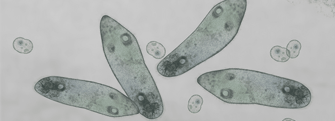
Microscopic image of paramecium and amoeba to represent live cell imaging techniques
Microscopy and Imaging | Lumenera

An Introduction to Live Cell Imaging

ByLumenera

Read on to learn more about live cell imaging, including how high rate microscopy can help you capture rapid cellular processes.

Read More An Introduction to Live Cell ImagingContinue

A gloved hand inserting PCR tubes into a thermocycler to represent answering the question how to get started with PCR
PCR, qPCR and qRT-PCR

What is PCR? The Beginner’s Guide

ByDr Jennifer Redig

If you need to copy, sequence, or quantify DNA, you need to know about PCR. Read our guide to the PCR process, and discover tips to help you avoid the most common PCR pitfalls.

Read More What is PCR? The Beginner’s GuideContinue

A stone staircase flanked by flowerpots to represent a step-by-step guide to qPCR primer design
PCR, qPCR and qRT-PCR

A Step-by-Step Guide to Designing qPCR Primers

ByNew England Biolabs

qPCR primer design is a bit of science, a bit of magic, and a little bit of luck. Here’s the science to help you design the best primers for your experiments.

Read More A Step-by-Step Guide to Designing qPCR PrimersContinue

Image of a colorful spring toy depicting coils in DNA plasmid preps to help undertand why you get 3 bands when running plasmid DNA on agarose gels
DNA / RNA Manipulation and Analysis

How to Identify Supercoils, Nicks and Circles in DNA Plasmid Preps using Gel Electrophoresis

ByDr Rebecca Tirabassi

Why do you get three bands when running uncut plasmid DNA on agarose gels. Discover the answer and how it can help improve your DNA plasmid preps.

Read More How to Identify Supercoils, Nicks and Circles in DNA Plasmid Preps using Gel ElectrophoresisContinue

Three shot glasses and a clear glass bottle containing clear alcoholic liquid to represent the ethanol grades that can be used in the lab
DNA / RNA Manipulation and Analysis

Which Grade of Ethanol Should I Use?

ByCristy Gelling

Want to know more about ethanol grades commonly used in the lab? We help you make sense of your flammables cabinet with our rundown of the ethanol grades typically used in molecular biology, as well as some important rules for how to use them correctly.

Read More Which Grade of Ethanol Should I Use?Continue

An unhappy looking man covered in soot and holding a cable with exposed wires to represent choosing between constant current or voltage in SDS-PAGE
Protein Expression and Analysis

Constant Current or Voltage in SDS-PAGE: The Great Debate

ByZandile Nare

It’s not always easy deciding whether to run electrophoresis at a constant voltage, current, or power. Here, we outline the differences to help you make an informed decision.

Read More Constant Current or Voltage in SDS-PAGE: The Great DebateContinue

Man holding a lens up to the light to represent lenses and objectives
Microscopy and Imaging

How History Shaped Modern Optical Microscopes, Part Two: Corrected Lenses and Objectives

ByMartin Wilson

Discover how chromatic and geometric imaging aberrations have been corrected over the last few centuries with the development of corrected lenses and objectives.

Read More How History Shaped Modern Optical Microscopes, Part Two: Corrected Lenses and ObjectivesContinue

A street sweeper sweeping leaves from a road to represent DNA clean-up
DNA / RNA Manipulation and Analysis

5 Ways to Clean Up A DNA Sample

ByDr Nick Oswald

Are you struggling with your DNA clean-up? Then check out our top five methods so you can pick the best option for your experiments.

Read More 5 Ways to Clean Up A DNA SampleContinue

Vintage microscope to represnet the history of simple and compound microscopes
Microscopy and Imaging

How History Shaped Modern Optical Microscopes, Part One: Simple and Compound Microscopes

ByMartin Wilson

Discover the history of simple and compound microscopes in this first of our two-part series on the history of microscopes.

Read More How History Shaped Modern Optical Microscopes, Part One: Simple and Compound MicroscopesContinue

Fingers "walking" up wooden blocks arranged as steps, representing the steps in Ct values from a qPCR experiment, analysed by double delta Ct analysis
PCR, qPCR and qRT-PCR

4 Easy Steps to Analyze Your qPCR Data Using Double Delta Ct Analysis

BySuganth Kannan

New to qPCR? Here’s a quick summary of one of the two most common analysis methods – double delta Ct analysis.

Read More 4 Easy Steps to Analyze Your qPCR Data Using Double Delta Ct AnalysisContinue

Image of a lumberjack cutting a tree to represent tissue sectioning tips
Microscopy and Imaging

Advanced Sectioning Techniques: How to Section Difficult Tissues

ByDr Jennifer Redig

Are you having problems with tissue sectioning? Follow these 10 tissue sectioning tips to create the perfect tissue section every time without stressing out.

Read More Advanced Sectioning Techniques: How to Section Difficult TissuesContinue

Image of vintage stereo as a play on words to represent a stereo microscope
Microscopy and Imaging

Stereo (Dissecting) Microscopes 101

BySteven Miller

Want to know about stereo microscopes? This article answers what a stereo microscope is, how it works and why it’s a great tool for biologists!

Read More Stereo (Dissecting) Microscopes 101Continue

Gloved hands of a researcher holding a Petri dish with green stained medium and surrounded by other stacked Petri dishes with red and yellow stains to represent someone identifying microbes
Cells and Model Organisms

Crash Course in Microbial Identification

ByDr. Karen O'Hanlon Cohrt

Need a crash course in microbial identification methods? Here we give you a rundown of the methods available for the identification of bacteria, yeast, or filamentous fungi to the species level.

Read More Crash Course in Microbial IdentificationContinue

multicoloured cells imaged on a confocal microscope to illustrate how you can improve your confocal images
Microscopy and Imaging

Confocal Images: Expert Tips for Your Hour of Need!

ByDr Nick Oswald

Getting publication perfect confocal images can be tricky. If you are struggling or just want to ensure you’re capturing the best images possible, check out our top 7 tips for confocal imaging.

Read More Confocal Images: Expert Tips for Your Hour of Need!Continue

light being defracted by a prism to represent optimizing the refractive index in confocal microscopy
Microscopy and Imaging

Resolution and Refractive Index: Set-up Your Confocal Wisely!

ByMelanie Laederich

Get the best out of your time on the microscope by understanding the refractive index of your experiment to optimize and increase your resolution.

Read More Resolution and Refractive Index: Set-up Your Confocal Wisely!Continue

Image of a iced coffee (Frappe) as a play on words for the microscopy technique FRAP
Microscopy and Imaging

Fun With FRAP! Fluorescence Recovery After Photobleaching for Confocal Microscopy

ByCatriona Paul

If you’re thinking FRAP is short for frappuccino then you need to read this article. Discover the history, how it works, and why you’d want it in your confocal toolbox

Read More Fun With FRAP! Fluorescence Recovery After Photobleaching for Confocal MicroscopyContinue

Two hands reach toward each other to connect chain links to represent various tricks for peptide synthesis
Protein Expression and Analysis

Synthetic Peptides Part 2: Tips and Tricks for Peptide Synthesis

ByHeinz Reiske

Deciding on the right peptide sequence can make or break your experiment. Find out what to keep in mind when designing a synthetic peptide.

Read More Synthetic Peptides Part 2: Tips and Tricks for Peptide SynthesisContinue

Colourful palms to represent Photoactivated Localization Microscopy (PALM).
Microscopy and Imaging | Zeiss

An introduction to Photoactivated Localization Microscopy (PALM)

ByZEISS Microscopy

How does photoactivated localization microscopy (PALM) work? And what use can PALM microscopy be to you? This short introduction to PALM gives you the answers!

Read More An introduction to Photoactivated Localization Microscopy (PALM)Continue

Image of a fluorescent flower in the palms of hands to represent Photoactivated Localization Microscopy and PALM sample prep
Microscopy and Imaging | Zeiss

Beginners Guide to PALM Sample Preparation

ByZEISS Microscopy

Good PALM sample preparation is the key to great images. Find out how to choose the right fluorescent proteins and learn some tips and tricks for sample prep.

Read More Beginners Guide to PALM Sample PreparationContinue

Image of a homemade digital microscope - the end product if you follow our instructions to build your own microscope
Microscopy and Imaging

Turn a Smartphone into a Digital Microscope for Only 3 Cents

ByMelissa Walker

Want to build your own microscope for almost nothing? You probably already have most of the tools that you need right in your lab or at home! Here’s how.

Read More Turn a Smartphone into a Digital Microscope for Only 3 CentsContinue

A agarose gel being loaded to represent tips for troubleshooting agarose gels given in the article
DNA / RNA Manipulation and Analysis

How to Get a Picture-Perfect Agarose Gel

ByHelena Mello

Check out our agarose gel hacks for troubleshooting your blurry or uneven bands, and tips for getting picture-perfect agarose gel images every time.

Read More How to Get a Picture-Perfect Agarose GelContinue

The tweezers used to hold the cherry are indicative of optical tweezers described in the article
Microscopy and Imaging

Trapping Objects With Light: From Star Wars to Neurons

ByFederico Iseppon

Can lasers be used to trap and move objects? Sure they can! Read on to know how you can use lasers in a cool technique called optical tweezers to manipulate minuscule objects under the microscope.

Read More Trapping Objects With Light: From Star Wars to NeuronsContinue

Image of person puzzled by a maze, indicating the complex nature of troubleshooting site-directed mutagenesis and the need for site-directed mutagenesis tips
PCR, qPCR and qRT-PCR

Troublesome Site-Directed Mutagenesis: Troubleshooting Your Experiment for Stubborn Mutations

ByLauren Tebay

As is sadly the case in many experiments, site-directed mutagenesis (SDM) does not always work the way we would like it to the first time around. Here are a few tips to help you on your way when trying to troubleshoot a bothersome SDM reaction!

Read More Troublesome Site-Directed Mutagenesis: Troubleshooting Your Experiment for Stubborn MutationsContinue

Image of 4 zebrafish to depicit how to measure movement in zebrafish
Cells and Model Organisms

Get a Move On: How to Measure Movement in Zebrafish

ByEmily O'Connor

If you are interested in the sensory or motor function of your zebrafish model, this is the test to try.

Read More Get a Move On: How to Measure Movement in ZebrafishContinue

Image of a fluorescent sign saying carpe diiem to represent the issue of photodamage in fluorescent live-cell imaging
Microscopy and Imaging

7 Steps to Avoid Photodamage in Fluorescent Live-cell Imaging

ByKimmo Tanhuanpää

Don’t get overexcited but we’ve got 7 top tips to help you minimize photodamage during your fluorescent live-cell imaging experiments.

Read More 7 Steps to Avoid Photodamage in Fluorescent Live-cell ImagingContinue

A representative electron micrograph image of axons ready to measure myelin thickness
Microscopy and Imaging

How to Measure Myelin Thickness Manually (and Other Tedious Lab Tasks) and Keep Your Sanity

ByNiamh McNamara

Tips on staying sane from a student survivor of tedious lab tasks.

Read More How to Measure Myelin Thickness Manually (and Other Tedious Lab Tasks) and Keep Your SanityContinue

Image of eggs, one with a helmet and another getting hit by a hammer to express the dos and don'ts of CAM assays
Cells and Model Organisms

6 Dos and Don’ts for Perfecting the CAM Assay

BySandesh Marathe

Don’t put all your eggs in one basket! Learn how to handle your eggs, prevent contamination and keep track of your experiments when performing the CAM assay.

Read More 6 Dos and Don’ts for Perfecting the CAM AssayContinue

A woman holding a magnifying glass representing detecting post-translational modifications
Protein Expression and Analysis

Detecting Post-Translational Modifications

ByAndrew Porterfield

A quick start guide to methods of assessing protein post-translational modifications

Read More Detecting Post-Translational ModificationsContinue

Image of cartoon chromosomes spread out on a plain background with a single chromosome in focusdepiciting preparing metaphase spreads
DNA / RNA Manipulation and Analysis

Preparing Metaphase Spreads: The Breakdown on Broken-Down Cells

ByKaitlyn Warren

A quick look at the first steps of metaphase spreads – the break down on breaking down your cells and the factors to keep in mind.

Read More Preparing Metaphase Spreads: The Breakdown on Broken-Down CellsContinue

Image of alphabet, depicting the A-Z of post-translational modifications
Protein Expression and Analysis

A-Z of Post-Translational Modifications

ByVicki Doronina

You might know the most common post-translational modifications, but there are many more than just phosphorylation and ubiquitination – come and test your knowledge!

Read More A-Z of Post-Translational ModificationsContinue

A man and a woman add a link to a growing chain to symbolize creating synthetic peptides
Protein Expression and Analysis

Synthetic Peptides and Their Uses

ByHeinz Reiske

Are you studying a small peptide or protein? Learn whether using synthetic peptides can save you hours of transfection, protein expression, and purification.

Read More Synthetic Peptides and Their UsesContinue

Image of garbage bags and recyling. Used as a metaphor for measuring autophagic flux
Protein Expression and Analysis

An Experimental Toolkit for Measuring Autophagic Flux

ByRebecca Wallings

How good is your cell at recycling? In this guide, we explain what autophagic flux is, why you need to study it, and methods to get you started.

Read More An Experimental Toolkit for Measuring Autophagic FluxContinue

Hand holding a brown egg to depict using chick embryps for studying angiogenesis
Cells and Model Organisms

Chick Embryo: A Model System for Studying Angiogenesis

BySandesh Marathe

Are you studying angiogenesis? Is cell culture not cutting it but you’re not ready for an in vivo model? Check out the CAM assay.

Read More Chick Embryo: A Model System for Studying AngiogenesisContinue

Laboratory mouse representing genetic and chemically induced mouse models
Cells and Model Organisms

Chemically Induced Mouse Models – Where Consistency Matters

ByGeil Merana

Tips and tricks to get the most optimum and consistent results in a DSS-induced colitis model

Read More Chemically Induced Mouse Models – Where Consistency MattersContinue

Red check mark in checklist to highlight things to consider when detecting non-histone lysine acetylation
Protein Expression and Analysis

Top 5 Things to Keep in Mind When Detecting Non-Histone Lysine Acetylation

ByVictory Ibigo Poloamina

Lysine acetylation isn’t just for histones. Read on to find out how to determine whether your favorite protein is lysine acetylated.

Read More Top 5 Things to Keep in Mind When Detecting Non-Histone Lysine AcetylationContinue

An assortment of paper gift tags to represent protein purification affinity tags
Protein Expression and Analysis

Recombinant Protein Expression: How to Choose the Right Affinity Tag

ByShubhranshu Zutshi

Using the right affinity tag can make protein purification a breeze. Read more to find out which tag is right for you.

Read More Recombinant Protein Expression: How to Choose the Right Affinity TagContinue

Image of Size Analysis of DNA using Gel Electrophoresis
Sage Science | Genomics and Epigenetics

Size Analysis of High-Molecular-Weight DNA for Long-Read Sequencing

BySage Science

Discover how to check DNA quality for long-read sequencing using electrophoresis and why pipetting carefully is so important.

Read More Size Analysis of High-Molecular-Weight DNA for Long-Read SequencingContinue

Moire wave to depicit how Structured Illumination Microscopy works
Microscopy and Imaging

Structured Illumination Microscopy (SIM) – An Introduction

ByDr Nick Oswald

Discover how structured illumination microscopy works can help you see things in greater detail.

Read More Structured Illumination Microscopy (SIM) – An IntroductionContinue

Scientist holding a blood smear slide with microscope in the foreground
Microscopy and Imaging

Making the Perfect Blood Smear

ByHaley Gassel

Stop wasting time throwing out slide after slide trying to create a readable blood smear. Read our how-to guide for creating the perfect blood smear slide.

Read More Making the Perfect Blood SmearContinue

Page navigation

Previous PagePrevious 1 … 3 4 5 6 7 … 31 Next PageNext
Accelerate your research career with curated daily lab wisdom

We collate wisdom and tools from researchers worldwide to help you to accelerate your progress.

Sign up now to get it in your inbox

Webinars

Podcasts

Newsletters

Articles

Downloads

About Us
  • About Us
  • Meet the Mentors
  • Get Involved
  • Contact Us
  • Get the T-Shirt
Education
  • Technical Skills
  • More Skills
  • Events
  • Podcasts
Marketing
  • Life Science Marketing
  • Community Marketing
  • Custom Marketing
Bitesize Bio Powered
  • Microscopy Focus
  • Privacy Policy
  • Cookie Policy
  • Terms of Use

Copyright © Science Squared – all rights reserved

10 Things Every Molecular Biologist Should Know

The eBook with top tips from our Researcher community.

Get The Tips
Scroll to top