Optimal Conditions for Live-Cell Imaging

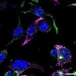
Live-cell imaging is the investigation of dynamic physiological processes in living cells using time-lapse imaging from milliseconds to hours. Live-cell imaging turns multiple snapshots to movies, which is in contrast to fixed-cell imaging that examines cellular activity at a time point. Typical applications of live-cell imaging that are used to study kinetic events include enzyme activity, signal transduction, protein trafficking, and membrane recycling process. Successful imaging relies on ensuring you achieve optimal conditions for live-cell imaging.
Performing Live-Cell Imaging on Cultured Cells: Media Choice
Live-cell imaging can be performed with cultured cell lines such as HEK cells and HeLa cells, primary cell cultures such as skin cells. You can even image whole brains inside living mice or rats, using a suitable brain imaging microscope.
Growth media or saline-based media formulations such as DMEM, RPMI, and Leibovitz L-15 are widely used in live-cell imaging experiments. The growth media solutions are formulated to support growth without components that affect background fluorescence.
On the other hand, saline formulations contribute to buffering and optical clarity, but cannot be used in long-term studies that demand high metabolic activity. Thus, proper functioning cells are important to study active biological processes during microscopy. But many cells cannot adapt to deviations from their optimal growth environments including pH, buffering capacity, temperature, O2 concentration, and osmolarity.
This article will cover the optimal conditions for live-cell imaging for maintenance of native state within cells.
Optimal Conditions for Live-Cell Imaging: Four Key Factors
pH
The pH value is critical for cell lines to grow efficiently. For instance, most cell lines propagate the best at pH 7.2-7.4, fibroblasts thrive up to pH 7.7, and transformed cell lines excel at around pH 7. To keep track of the changes in media pH during cell growth phases, phenol red is used in some cases as an indicator of pH value, which is red at pH 7.4. The phenol red turns into orange (pH 7) and yellow (pH 6.5) in acidic solutions, and becomes pink (pH 7.6) and purple (pH 7.8) in basic solutions. However, common media formulations in live-cell imaging avoid phenol red due to its high extinction coefficient of light absorption which may contribute to background noise.
Buffers
The pH-buffering system typically contains sodium bicarbonate and an external supply of CO2. In addition, synthetic biological buffers such as HEPES may be applied to buffer the media in short-term studies. When extracellular solutions cannot be buffered with HEPES, CO2 is delivered to the system which dissociates into bicarbonate when in contact with the extracellular solutions. That’s why live-cell imaging on the microscope usually requires incubation chambers that are specifically designed for a regulated atmosphere. In general, a concentration of 10-20mM HEPES buffer can control pH in the absence of CO2 atmosphere, with sodium bicarbonate provided for optimal growth.
O2 Concentration
Cell lines can vary widely in their O2 requirement. Mammalian cells usually require oxygen for respiration in vivo, but can often substitute anaerobic glycolysis when grown in vitro as primary lines or after immortalization. But strict O2 regulation is not necessary for most live-cell imaging experiments. Interestingly, depletion of oxygen is often used as a strategy to reduce the photo-damage that can occur through reactions with reactive oxygen species. This strategy may involve either an O2 depletion system such as oxyrase or antioxidants such as vitamin C to limit the free radical damage. However, reduced O2 tension may result in hypoxic stress in some cases, which can be dangerous to cells.
Osmolarity
Most cell lines grow efficiently at a wide range of osmolarity value between 260 and 320 mosM. It is important to monitor osmolarity of the culture medium when altering the constitution by the addition of HEPES. Small volumes of media accommodated by most imaging chambers are subject to changes in osmolarity due to evaporation when the temperature of medium is high. In most cases, microscopes have a CO2-controlled incubation chamber for working with mammalian cells at 37oC. The hypotonic medium can be routinely substituted in Petri dishes to compensate for evaporation. Evaporation can also be minimized either by sealing the medium in an open chamber with oil or by controlling humidity.
Table 1. Overview of environmental control for live imaging of mammalian cells. Adapted from Nikon Instruments Inc.[1]
Parameters | Optimum Range |
---|---|
pH | 7.0-7.7 |
Buffer | Sodium bicarbonate or Synthetic biological buffers |
Temperature | 28-37°C |
Osmolarity | 260-320 mosM |
Atmosphere | Air or 5-7% CO2 |
Humidity | 97-100% |
Oxygenation | Variable |
Tips for Achieving Optimal Conditions for Live-Cell Imaging
The application of large volume of medium in short-term studies may avoid deviations in osmolarity and O2 due to evaporation. For long-term studies, a combination of heating units, humidified incubation chamber, and bicarbonate-based buffering system can be applied to control the environmental parameters.
Often advanced widefield and confocal microscopy techniques are applied for the better interpretation of results in live-cell imaging experiments. For example, cellular growth and development are observed under phase contrast and differential interference contrast (DIC) microscopy but developmental embryonic studies are sometimes preferred to observe under stereo microscopy. On the other hand, fluorescent techniques are used to label specific cellular compounds which make them better observable under confocal microscopy.
However, live-cell imaging systems are limited to certain optimal conditions to make sure the cells are maintained in a healthy state. Taken together, live-cell imaging requires both focusing on cells functioning during the experiment and applying specific experimental methods. Thus it is important to review the application of media, environmental control, pH, and imaging techniques in such experiments where a minimal adjustment may change the cellular processes of interest.
Do you want more information on live-cell imaging? Find out how to pick the right live-cell imaging technique for your research or leave us a comment below.
References
- Nikon. Maintaining Live Cells on the Microscope Stage. Accessed May 2021.
- Thermo Fisher Scientific. Preparing for Live-Cell Imaging. Accessed May 2021.
- Leica Microsystems. Live Imaging. Accessed May 2021.
Originally published January 16, 2019. Reviewed and updated May 2021.