Having some knowledge of microscope history can be beneficial to understanding and appreciating specific configurations and components, and how they produce an optimal image. For example, practically all laboratory microscopes use achromatic objectives to partly compensate for chromatic aberration. For high-quality imaging, fluorescence microscopy, and photomicroscopy, fluorite, and apochromatic objectives are used to largely overcome chromatic and geometric aberrations.
The three microscope configurations you are likely to use include the stereo, inverted microscope, and compound microscope. This covers a vast range, from routine instruments to research-grade microscopes forming part of a complete imaging system. This article traces the brief history of these instruments and their main components, the developmental problems encountered, and the innovations we rely upon.
What Do We Mean By Simple and Compound Microscopes?
Here we will explore the brief history of early microscopes, focusing on simple and compound microscopes.
Simple Microscopes
Simple microscopes consist of a single lens mounted in a holder that is either fixed, or can be adjusted for focusing. Although the invention of this microscope dates back to the 17th century, single-lens instruments were widely manufactured in the late 1800s and early to mid-1900s for use as dissecting microscopes. Simple microscopes are gaining popularity again with 2D/3D folded and printed versions that are easily constructed, providing an inexpensive instrument that can be used in harsh environments such as disease identification in disaster or emergency relief situations.
Choose a free resource to help you move forward
POSTER
Fluorescent Proteins Guide
EBOOK
Guide to Special Stains for Histology
Compound Microscopes
The compound microscope derives its name as overall magnification is achieved using two main optical components: (1) the objective lens and (2) the eye-piece (or ‘ocular’). The magnification of the objective lens is multiplied, or ‘compounded’ by the eye-piece. The modern compound microscope typically consists of a number of objectives (mounted on a ‘nosepiece’ or turret) with either monocular or binocular eyepieces.
A Five-Year-Old Inventor?
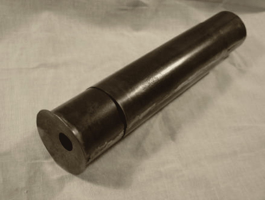
The compound microscope was created and developed between the 16th and 17th centuries. It is generally accepted that Zaccharias Janssen (1585-1632), an eyeglass maker from the Netherlands, is the inventor, but he was five to ten years old at the time of invention (around 1590 to 1595). One theory is that he helped his father in their workshop and the credit should go to them both. Their microscope consisted of a handheld central cylinder with moveable eyepiece and objective tubes (Figure 1). The bi-convex eyepiece and plano-convex objective lenses produced a magnification of between three and nine times.
Another claimant to the invention is Hans Lippershey (1570-1619), a German eyeglass maker. Although historians agree that Lippershey invented the telescope, there is debate as to whether he independently invented a microscope. Indeed, some accounts claim he stole the idea from Janssen and further accounts claim he stole the idea for the telescope!
Naming the Instrument
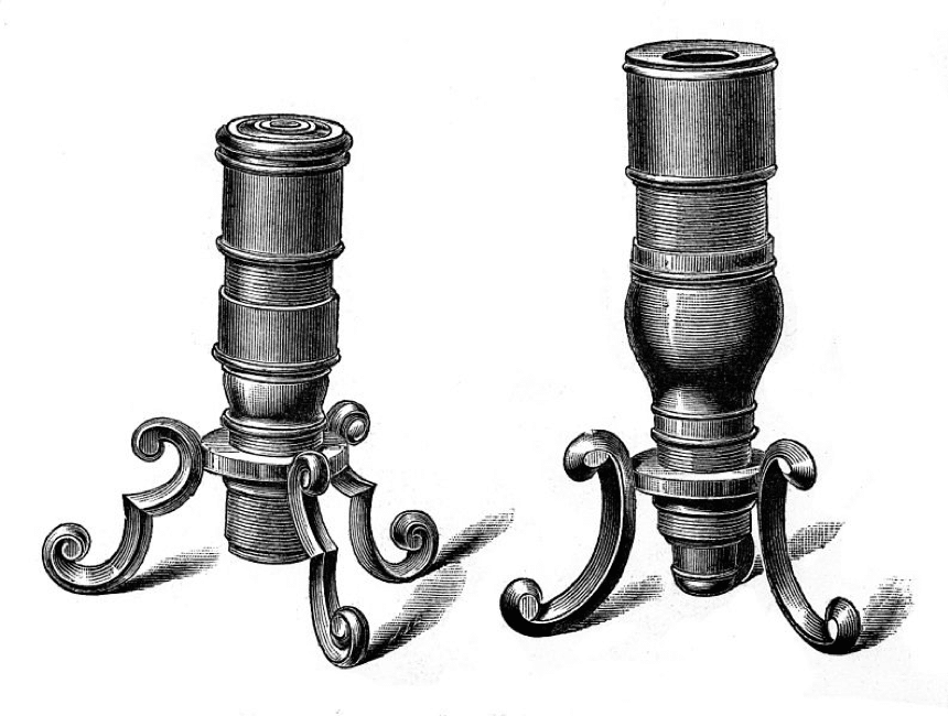
News of the invention spread across Europe, coming to the attention of Galileo Galilei (1564-1642) and his friend, the botanist Giovanni Faber (1574-1629). Galileo built his ‘occhiolino’ in 1609, and Faber was the first to name the ‘microscope’ from the Greek ‘micron’ (small) and ‘skopein’ (to look at). Essentially a modified telescope, Galileo’s microscope used a bi-concave eyepiece and bi-convex objective lens to provide up to 30 times magnification. Although none of Galileo’s microscopes survive, his creations featured a tripod stand for vertical specimen viewing (Figure 2).
Micrographia
The next major development came with the publication of ‘Micrographia’ in 1665 by the English natural philosopher Robert Hooke (1635-1703). This first illustrated microscopy book contained detailed engravings including the eye of a fly and a complete flea (Figure 3). Hooke’s microscope was made by Christopher Cock of London and featured a bi-convex objective and eye lenses, and a field lens for increased field of view. However, in practice, this lens decreased resolution and Hooke removed it to examine the finer details of specimens. Hooke’s design was one of the first to feature a stand allowing height and angle adjustment. In addition, he used an oil lamp and water-filled globe to concentrate light on his samples, essentially acting as a condenser. Finally, Hooke is famous for discovering and naming the cell, following his observations of a cork sample.

The Father of Microbiology
Microscope development wouldn’t be complete without mentioning Antonie van Leeuwenhoek (1632-1723), who made around 500 instruments and lenses (though only 10 of these exist today). His design differed from other instruments of the time, consisting of a single bi-convex lens between two metal plates (Figure 4). His ability to grind and polish extremely high-quality lenses resulted in instruments producing up to 275X magnification. Furthermore, these simple microscopes didn’t suffer from aberrations caused by combined lens designs used by his predecessors. Through his work, van Leeuwenhoek discovered protozoa and bacteria, and he is known as the ‘Father of Microbiology’.
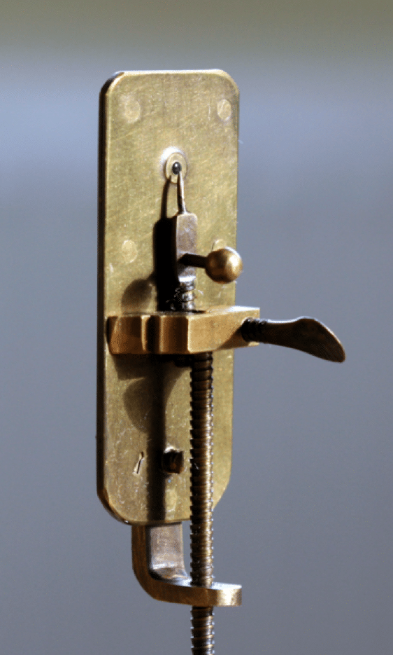
Table 1: Summary of dates, inventors, and their microscopes.
| Date | Inventor/Maker | Microscope type | Magnification |
| 1590 to 1595 | Hans and Zacharias Janssen | Compound (bi-convex eyepiece/plano-convex objective) | 3X to 9X |
| 1609 | Galileo Galilei | Compound (bi-concave eyepiece/bi-convex objective) | Up to 30X |
| Late 1600s | Robert Hooke/Christopher Cock | Compound (bi-convex eyepiece and objective with removable field lens) | Up to 50X |
| 1676 | Antonie van Leeuwenhoek | Simple (single bi-convex lens) | 70X to 275X |
Corrected Lenses and Objectives
As mentioned, laboratory microscopes rely upon objectives corrected for chromatic and geometric aberrations. The history of corrected lenses and objectives is vast, with numerous pioneers, but this next section provides a general overview of these imaging anomalies and the developments made over the last few centuries.
Geometric and Chromatic Aberrations
Early microscope lenses were low quality and of imperfect shape, producing sub-optimal imaging as a result of geometric and chromatic aberrations.
There are five geometric (or ‘Seidel’) aberrations: (1) astigmatism; (2) coma; (3) distortion; (4) spherical; and (5) field curvature. For the purpose of brevity, this article focuses on the development of lenses corrected for spherical aberration.
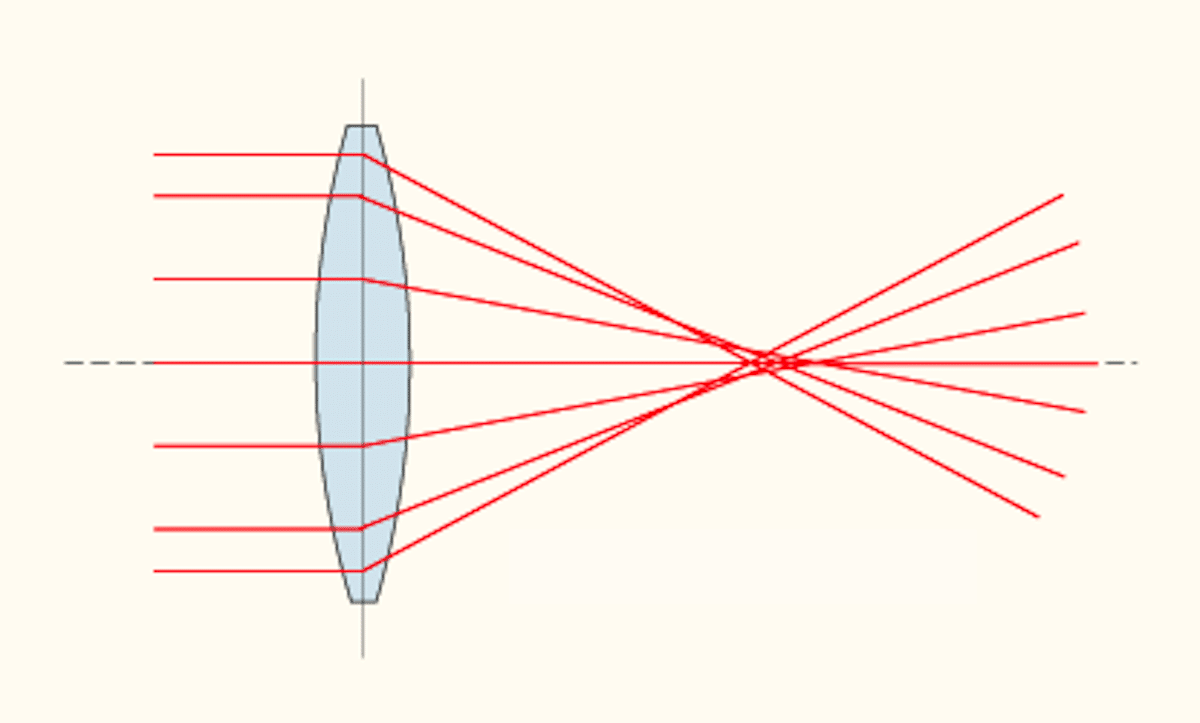
In brief, spherical aberration is related to the structure and geometry of the lens and the refraction of light as it passes through it. Light waves passing near the edge of a curved lens are refracted to a greater extent than those at the center. Focal points are therefore spread out along the optical axis, with blurred imaging and reduced resolution (Figure 5).
Chromatic aberrations are related to the lens material, which may cause white light to be split into constituent wavelengths. The refractive index of a lens varies according to wavelength, and with a white light source, the red component is refracted the least, followed by green, and then blue. As a result, constituent colors don’t converge at the same focal point, resulting in colored fringes or halos (Figure 6).

Objectives corrected for the aberrations mentioned above are termed ‘achromatic’, ‘apochromatic’, and ‘fluorite’, with the differences highlighted below (Table 1).
| Objective | Chromatic aberration correction | Spherical aberration correction | Abbreviated name (engraved on objective barrel) |
| Achromatic (or ‘Achromat’) | Blue and red | Green | ‘Achro’ or ‘Achromat’ |
| Fluorite (or Semi-Apochromatic) | Blue and red | Two to three wavelengths | ‘Fl’, ‘Fluo’, ‘Fluar’ or ‘Fluor’ |
| Apochromatic (or ‘Apochromat’) | Blue, green and red | Two to three wavelengths | ‘Apo’ |
The First Achromatic Lenses
The first achromatic lens was developed around 1729 by the English mathematician Chester Moore Hall (1703-1771). By fusing crown glass and flint glass, Hall used this lens to build an achromatic telescope in 1733.
Despite Hall’s work, the English optician John Dollond (1706-1761) is credited as the inventor of the achromatic lens because he was the first to patent the design. As a professional courtesy to Hall’s earlier developments, Dolland did not enforce the patent. However, following John’s death, his son Peter Dollond (1730-1820) enforced the patent, leading to controversy and bitter court battles with 35 other opticians. Nevertheless, these lenses were large and unsuitable for microscopy.
The Development of Achromatic Objectives
Another 50 years passed until the achromatic lens was applied to the field of microscopy. This was achieved by the father and son team of Jan (1715-1801) and Harmanus van Deyl (1738-1809). In 1770, they created lenses small enough for a microscope objective, and were the first to commercially offer their creation in 1807. Their objectives provided a resolution of 5 \(mu\)m with a compounded magnification up to 150X.
Further achromat refinements were made by the German glass and mirror maker, Joseph von Fraunhofer (1787-1826). His greatest contributions to optical theory are highlighted in his work on the solar spectrum (the Fraunhofer spectral absorption lines were named in his honor). By improving the quality of optical glass, and leaving an air gap between objectives lenses, he reduced chromatic aberration. This gap configuration (with some refinements) is still used today.
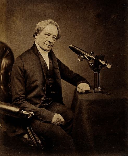
Whilst improved achromats were developed in the 18th and 19th Centuries, spherical aberration continued to hamper optimal imaging, and early attempts to correct this decreased specimen brightness and resolution. Despite working as a wine merchant, Joseph Jackson Lister (1786-1869; father of Joseph Lister, who pioneered antiseptic surgery) had a very keen interest in optics and microscopy (Figure 7). Lister began studying lenses in the mid-1820s, discovering that varying the distance between lenses could reduce aberrations. Lister published a paper on improved lenses in 1830 and collaborated with Andrew Ross (1798-1859), a renowned London microscope maker. Using Lister’s designs, Ross constructed improved achromatic lenses that were chromatically corrected for two wavelengths and spherically corrected for one. Lister and Ross were two of the 17 people who founded the Microscopical Society of London in 1839 (now the Royal Microscopical Society).
Ernst Abbe- His Theories and Applications
At this point in history, lens correction had been a largely empirical pursuit, but this was about to change with the work of Ernst Abbe (1840-1905). Abbe proposed many theories and formulae that still form a substantial basis for lens design and imaging, including the designation of numerical aperture.
In 1866, Abbe was hired by Carl Zeiss (1816-1888), who had been making and selling microscopes since 1847. Zeiss was aware of the competition and that optical component production had relied on trial-and-error. He aimed to produce instruments “determined beforehand by computation” and appointed Abbe as Research Director of the Zeiss Optical Works. From 1872, all Zeiss microscopes were produced according to Abbe’s theories.
However, with the quality of glass available, objectives exhibited variations in refractive index leading to sub-optimal resolution and imaging. In 1879, Abbe received a new lithium glass sample from the chemist Otto Schott (1851-1935). Abbe brought Schott to the German city of Jena in 1882, where a glass-making laboratory was set up for him. They collaborated with Zeiss to form a business, now known as ‘Schott’ (famous for Duran glassware).
Around 1881, Abbe began experimenting with the mineral fluorite, postulating that its optical properties could reduce chromatic and spherical aberrations. After receiving fluorite from the Oltscheren Alps, Abbe traveled to the region in 1886 and spent time at the hut owned by Zeiss Optical Works. Fluorite may have been used in the first apochromatic objectives, although this was never detailed.
In 1886, the company produced the most precise objective, the apochromat, eliminating chromatic aberration for three wavelengths and largely minimizing spherical aberration. Despite initial lens problems, Zeiss Optical Works paved the way for an objective that was to revolutionize microscopy, forming the basis of modern apochromatic objectives.
Abbe published his work on fluorite in 1890, and this objective provided greater spherical aberration corrections than the achromat. The fluorites were (and still are) more affordable than the apochromat, and although some modern lenses use a synthetic substitute, the mineral is still used in objectives.
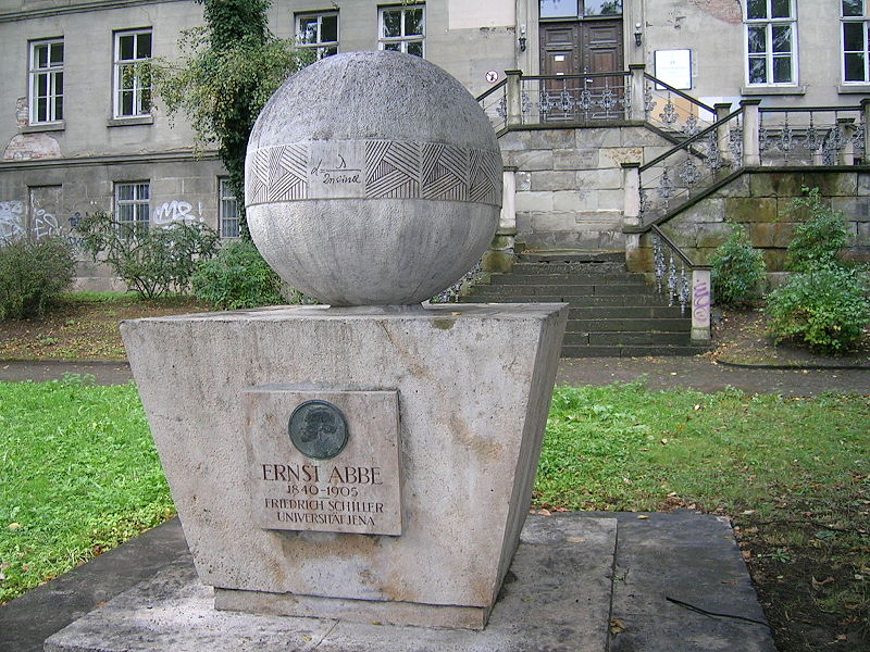
In 1889, a year after Zeiss died, Abbe established the Carl Zeiss Foundation in his honor. He also transformed conditions for the employees, including the introduction of an eight-hour day. Abbe retired in 1903, and a torchlight procession of 1500 Zeiss employees was held in his honor. He died in 1905 and is remembered in Jena in the form of two memorials (Figure 8).
Optimal microscope imaging was largely hindered until the latter half of the nineteenth century due to the inherent imperfections of early lenses combined with the trial-and-error approach to lens correction. Without detracting from the work of the early pioneers, we have Ernst Abbe to thank for the basis of imaging theory and modern lens design.
The timeline below summarises the development of corrected lenses and objectives.
| Date | Inventor/Maker | Component | Notes |
| 1729 | Chester Moore Hall | Achromatic lens | Crown and flint glass lens, for telescopes only. Hall built the first achromatic telescope in 1733. |
| 1758 | John Dollond | Achromatic lens | Patented the achromatic lens, but only suitable for telescopes. |
| 1770 | Jan and Harmanus van Deyl | Achromatic objective | Created the first commercially available achromatic objective microscope in 1807. |
| 1817 | Joseph von Fraunhofer | Achromatic objective | Vast improvements in glass and optical technology that led to reduction in chromatic aberration. |
| 1830 | Joseph Jackson Lister and Andrew Ross | Achromatic objective (two plano-convex achromatic doublets in aplanatic arrangement). | Objectives corrected for chromatic and spherical aberrations. |
| 1886 | Ernst Abbe, Otto Schott and Carl Zeiss | Apochromatic objective and Fluorite objective. | First objectives based on theoretical foundations. |
Final thoughts
Numerous inventors contributed to microscope development, but covering them all is out of the scope (pardon the pun!) of this article; therefore, please see the references and suggested reading below.
References
- Bradbury S. (1967) The Evolution of the Microscope. Pergamon Press, Oxford.
- Clark, E. Pioneers in Optics: Galileo Galilei. Microscopy Today. 2018;26(3): 48-49. DOI: https://doi.org/10.1017/S1551929518000470
- Davidson, M.W. Pioneers in Optics: John Dollond. Microscopy Today. 2016;24(2): 52-53.
- Davidson, M.W. Pioneers in Optics: Joseph von Fraunhofer and Gustav Robert Kirchhoff. Microscopy Today. 2011;19(5): 54-56.
- Davidson, M.W. Pioneers in Optics: Joseph Jackson Lister and Maksymilian Pluta. Microscopy Today. 2011;19(3): 54-56.
- Davidson, M.W. Pioneers in Optics: Ernst Abbe. Microscopy Today. 2009;17(1): 38-41.
- Jobst, R. The 120th anniversary of the Carl Zeiss Works, Jena, and the 150th anniversary of the birth of Carl Zeiss. Soviet Astronomy. 1967;11(1): 173-177;
- Lawson I. Crafting the microworld: how Robert Hooke constructed knowledge about small things. Notes and Records, The Royal Society Journal of the History of Science. 2016; 70 (1): 23-44. DOI: 10.1098/rsnr.2015.0057
- Lister J.J. (1830) On some properties in achromatic object-glasses applicable to the improvement of the microscope. Philosophical Transactions of the Royal Society of London, 120: 187-200.
- Robertson L.A. van Leeuwenhoek microscopes—where are they now? FEMS Microbiology Letters. 2015; 362 (9). DOI: https://doi.org/10.1093/femsle/fnv056
- Schott’s Glass- a ‘Chemistry World’ article from The Royal Society of Chemistry on Otto Schott’s work with Abbe and Zeiss.
- Von Rohr, M. The first Jena Catalog of Optical Glasses was published in 1886. Current Science. 1936;5(1): 25-30
Further Reading
- Further information on aberrations and modern corrected objectives. Leica Microsystems.
- A website dedicated to the life and work of Antonie van Leeuwenhoek, also containing information and images regarding Galileo’s and Hooke’s microscopes.
- An optics timeline, from prehistory to the 21st Century.
- An in-depth (400 page) illustrated article on the Microscopy and Histology collection at the Department of Cell Biology, Yale School of Medicine
- Wollman A. J. M., et al. From Animaculum to single molecules: 300 years of the light microscope. Open Biology. 2015; 5 (4). DOI: https://doi.org/10.1098/rsob.150019
You made it to the end—nice work! If you’re the kind of scientist who likes figuring things out without wasting half a day on trial and error, you’ll love our newsletter. Get 3 quick reads a week, packed with hard-won lab wisdom. Join FREE here.