Skip to content
Search
Technical Skills
Soft Skills
Events
Podcasts
Resources
Get Involved

About Us

  • About Us
  • Meet the Mentors
  • Get Involved
  • Contact Us
  • Get the T-Shirt

Marketing

  • Life Science Marketing
  • Community Marketing
  • Custom Marketing

Join Us
Sign up for our feature-packed newsletter today to ensure you get the latest expert help and advice to level up your lab work.

Sign Up now
  • Technical Skills
    • CRISPR
    • Histology
    • Lab DIY
    • Genomics & Epigenetics
    • DNA / RNA Manipulation and Analysis
    • Protein Expression & Analysis
    • PCR & Real-time PCR
    • Flow Cytometry
    • Microscopy & Imaging
    • Cells and Model Organisms
    • Analytical Chemistry and Chromatography Techniques
    • Chemistry for Biologists
    • Basic Lab Skills & Know-how
    • Equipment Mastery & Hacks
  • More Skills
    • Managing the Scientific Literature
    • Career Development and Networking
    • Dealing with Fellow Scientists
    • Getting Funded
    • Lab Safety
    • Lab Statistics & Math
    • Organization & Productivity
    • Personal Development
    • PhD Survival
    • Soft Skills & Tools
    • Software & Online Tools
    • Survive & Thrive
    • Taming the Literature
    • Writing, Publishing & Presenting
  • Events
  • Podcasts
  • Resources
  • Get Involved

Microscopy and Imaging

Vivid blue shapes on a dark background to show working positive controls for immunofluorescence
Microscopy and Imaging

5 Controls for Immunofluorescence: A Beginner’s Guide 

ByGauravi Deshpande

Achieving publication-quality immunofluorescence images is tricky. Learn what controls for immunofluorescence you can use to get them!

Read More 5 Controls for Immunofluorescence: A Beginner’s Guide Continue

Child using an abbacus to represent automatic cell counting
Microscopy and Imaging

Quick and Easy Automatic Cell Counting in 4 Steps

ByThomas Warwick

This article explains a simple, 4-step method for automatic cell counting with ImageJ. Perfect for your cell proliferation studies, gene expression analysis, or whatever your downstream application might be.

Read More Quick and Easy Automatic Cell Counting in 4 StepsContinue

Image of toilet tissue as a pun about tissue processing for histology
Microscopy and Imaging

Tissue Processing For Histology: What Exactly Happens?

ByMartin Wilson

Tissue processing for histology is a key step between fixation and embedding. We take you through the steps of tissue processing in this simple guide.

Read More Tissue Processing For Histology: What Exactly Happens?Continue

A researcher examining a microscope slide to fix adherent cells in a laboratory setting.
Microscopy and Imaging

How To Fix Adherent Cells For Microscopy And Imaging

ByDr Jennifer Redig

Adherent cell fixation is a crucial step in preparing cells for microscopy and imaging, ensuring that cellular structures are preserved for detailed analysis. Read our 8-step guide on how to effectively fix adherent cells to your microscope slides, including tips on sterilization, coating, and fixation methods, right here.

Read More How To Fix Adherent Cells For Microscopy And ImagingContinue

A polished cross-section of intricate rock as an abstract representation of the result of histology slide preparation
Microscopy and Imaging

How Histology Slides Are Prepared

ByNicola Parry

Ever wondered what magic happens to turn your samples into histology slides? Find out the 5 simple steps for histology slide preparation.

Read More How Histology Slides Are PreparedContinue

Abstract graphic of teal and orange shapes depicting the power and benefits of Volume Electron Microscopy.
Microscopy and Imaging

What is Volume Electron Microscopy and Should You Use it?

ByThomas Warwick

The emergence of Volume Electron Microscopy (vEM) has unlocked new possibilities in biological imaging, enabling us to visualize 3D structures of cells at high resolution. Learn more about this incredible technique in our latest article.

Read More What is Volume Electron Microscopy and Should You Use it?Continue

Abstract intricate represent hematoxylin and eosin staining.
Microscopy and Imaging

A Beginner’s Guide to Hematoxylin and Eosin Staining

ByNicola Parry

Discover what hematoxylin and eosin staining is used for and how it works, in this concise guide.

Read More A Beginner’s Guide to Hematoxylin and Eosin StainingContinue

Electron Microscopy Sample Preparation: 10 Pieces of Expert Advice
Leica | Microscopy and Imaging

Electron Microscopy Sample Preparation: 10 Pieces of Expert Advice

ByLeica Microsystems

Do you prepare samples for electron microscopy and want to save time, money, effort, and frustration? This article provides hands-on advice to help you get the best possible data out of your EM experiments.

Read More Electron Microscopy Sample Preparation: 10 Pieces of Expert AdviceContinue

Abstract lines of color to represent fluorescence from sample in confocal laser scanning microscopy
Microscopy and Imaging

Confocal Laser Scanning Microscopy Explained In 3 Easy Steps

ByCatriona Paul

Learn how confocal laser scanning microscopy works, its applications, and why it’s great for samples that are too thin to section.

Read More Confocal Laser Scanning Microscopy Explained In 3 Easy StepsContinue

A fluorescent fish as a pun on in situ hybridization
Microscopy and Imaging

How to Totally Nail Your First in situ Hybridization

ByRebecca Roberts

Having problems with your in situ hybridization? We’ve got 7 simple tips to help you get outstanding results.

Read More How to Totally Nail Your First in situ HybridizationContinue

Crayons to represent colors in digital images
Microscopy and Imaging

The 2 Types of Digital Images: Tools to Prepare Stunning Images for Publication

ByDaphne Ng

Digital images are essential to communicate your data. Get the information and tools you need to take your scientific illustration to the next level!

Read More The 2 Types of Digital Images: Tools to Prepare Stunning Images for PublicationContinue

Pins on a map to represent spatial separation in the context of protein colocalization.
Microscopy and Imaging

Protein Colocalization: 2 Essential Methods to Prove Protein Overlap

ByJelena Jankovic

You know the drill. To prove your theory, you must show the colocalization of X and Y in a cell. Here are 2 ways to reveal protein colocalization.

Read More Protein Colocalization: 2 Essential Methods to Prove Protein OverlapContinue

Picture of a fluoresence microscope set up
Microscopy and Imaging

Fluorescence Microscopy: From Principles to Applications

ByCamilla S. Colding-Christensen

Whether you want to get started with fluorescence microscopy or already use it, this guide will ensure you know the basics and get the best out of your fluorescence microscopy.

Read More Fluorescence Microscopy: From Principles to ApplicationsContinue

Two colourful springs representing the donor and acceptor in how FRET works
Microscopy and Imaging

How FRET Works: A Guide to Visualizing Protein Interactions

ByDr Nick Oswald

Not sure what FRET is, or just need a refresher on how FRET works? Read our short guide to understand the usefulness of FRET for studying protein-protein interactions.

Read More How FRET Works: A Guide to Visualizing Protein InteractionsContinue

Oil droplets to depict the use of oil in oil immersion microscopy
Microscopy and Imaging

How Using Oil Immersion Microscopy Can Increase Your Resolution

ByMartin Wilson

Oil immersion microscopy can improve your resolution in microscopy. This article will explain why this is the case and how you can use oil immersion microscopy in the lab!

Read More How Using Oil Immersion Microscopy Can Increase Your ResolutionContinue

A graphic of a fist punching the air to describe the concept of what is Cryo-Electron microscopy
Microscopy and Imaging

What Is Cryo-Electron Microscopy? A Brief Introduction

ByThomas Warwick

You don’t have to be a genius to understand Cryo-EM. Discover the fundamentals of this powerful microscopy tool and what propelled it into the scientific mainstream.

Read More What Is Cryo-Electron Microscopy? A Brief IntroductionContinue

An image of parchment and quill pen to depict the history of Cryo-Electron Microscopy
Microscopy and Imaging

A Short History of Cryo-Electron Microscopy

ByThomas Warwick

The slow, inching progress of cryo-EM towards the scientific mainstream can be told as a story with three parts. So take a step back and enjoy a short history of cryo-electron microscopy.

Read More A Short History of Cryo-Electron MicroscopyContinue

Explorer in a cave of ice to represent the extreme temperatures required during cryo-EM sample prep
Microscopy and Imaging

Cryo-EM Sample Prep: 5 Crucial Considerations 

ByThomas Warwick

You don’t have to be a brainbox to get your samples ready for cryo-EM, but a little wisdom goes a long way. Learn how to tend to your tissues, organize your organelles, and prepare your proteins to get the micrographs you’ve always dreamed of.

Read More Cryo-EM Sample Prep: 5 Crucial Considerations Continue

Little biomolecules in a magnifying glass showing the types of things that can be viewed using electron microscopy
Microscopy and Imaging

The 2 Main Electron Microscopy Techniques: SEM vs TEM

ByLouise Hughes

Microscopy is a huge and active field. Sometimes, it’s easy to forget the basics. Read our biologists’ guide to electron microscopy techniques.

Read More The 2 Main Electron Microscopy Techniques: SEM vs TEMContinue

Little red viruses to show biological applications of electron microscopy
Microscopy and Imaging

Applications of Electron Microscopy: An Easy Intro for Biologists

ByLiselle Bovell

Microscopy is a huge and active field. Sometimes, it’s easy to forget the basics. Read our biologists’ intro to applications of electron microscopy.

Read More Applications of Electron Microscopy: An Easy Intro for BiologistsContinue

An image of microscope that resembles a happy face to represent happy researchers following SEM sample preparation
Microscopy and Imaging

Scanning Electron Microscopy: 6 SEM Sample Preparation Pointers for Successful Imaging

BySaraswathi Sadasivan

Discover 6 critical scanning electron microscopy sample preparation points you need to know to get the best out of your SEM.

Read More Scanning Electron Microscopy: 6 SEM Sample Preparation Pointers for Successful ImagingContinue

Image of coloured dots to represent vizualizing RNA with light-up RNA aptamers.
Microscopy and Imaging

Light-up RNA Aptamers: Illuminating the World of RNA

ByVicki Doronina

Discover how you can visualize that notoriously difficult molecule, RNA using light-up RNA aptamers (LURAs).

Read More Light-up RNA Aptamers: Illuminating the World of RNAContinue

image of fluorescent lights to represent choosing a fluorescent protein
Microscopy and Imaging

The Ultimate Guide to Choosing a Fluorescent Protein

ByAdrienne Huntress

Discover the critical considerations when choosing a fluorescent protein, the key features of those most commonly used, and why newer might be better.

Read More The Ultimate Guide to Choosing a Fluorescent ProteinContinue

Image of a mechanic thinking before fixing a car to represent thinking before performing histology fixation
Microscopy and Imaging

An Introduction To Fixation For Histology: Think Before You Fix!

ByMike Millar

How you fix your tissue or cells can affect your results, for better or for worse. Discover the key points to think about before undertaking your histology fixation.

Read More An Introduction To Fixation For Histology: Think Before You Fix!Continue

A bookshelf of old books to represent the history of histology
Microscopy and Imaging

A (very) Short History of Histology

ByMartin Wilson

Discover the history of histology, from the first mention of a cell in 1665 to the identification and development of various stains.

Read More A (very) Short History of HistologyContinue

Image of a girl surround by traps to represent avoiding histology mistakes
Microscopy and Imaging

Avoid These Pitfalls: Seven (Not So Deadly) Histology Sins

ByDr Jennifer Redig

Discover seven common histology mistakes and how you can avoid making them when performing your experiments.

Read More Avoid These Pitfalls: Seven (Not So Deadly) Histology SinsContinue

Image of oil and water to represent understanding the importance of the numerical aperture on a microscope objective
Microscopy and Imaging

That Other Number – The Meaning Of Numerical Aperture In Microscopy

ByCynthia Barber

Do you know what that NA number is on your objective? We walk you through what the numerical aperture is and why it’s important.

Read More That Other Number – The Meaning Of Numerical Aperture In MicroscopyContinue

An image of microscope objectives with a scientist's hands to represent common microscope objective abbreviations
Microscopy and Imaging

An Objective View: What do These Abbreviations Mean on Microscope Objectives?

ByMartin Wilson

There are a large number of microscope objective abbreviations relating to optical aberrations; here we’ll shed some light on some of the most common ones to get you up to speed in no time!

Read More An Objective View: What do These Abbreviations Mean on Microscope Objectives?Continue

Image of 'pac man' style character eating red dots to represent live imaging of phagocytosis
Microscopy and Imaging

Live Imaging of Phagocytosis: Your Simple Guide to Capturing the Action

ByJude Bain

Live imaging of phagocytosis helps capture the details of this dynamic process. Discover tips and tricks to visualizing this important cellular process.

Read More Live Imaging of Phagocytosis: Your Simple Guide to Capturing the ActionContinue

Image of a microscope and a computer showing the sample image to represent an explanation of the point spread function
Microscopy and Imaging | Zeiss

A Beginner’s Guide to The Point Spread Function

ByZEISS Microscopy

Learn how the Point Spread Function affects what you see through your microscope and discover what you can do to improve your images.

Read More A Beginner’s Guide to The Point Spread FunctionContinue

An image of setting up a photography shoot to represent getting optimal conditions for live-cell imaging
Microscopy and Imaging

Optimal Conditions for Live-Cell Imaging

BySulov Saha

Live-cell imaging can bring a lot of clarity to cellular processes, but keeping your cells happy can be tricky. Read on to learn about 4 key parameters for achieving optimal conditions for live-cell imaging.

Read More Optimal Conditions for Live-Cell ImagingContinue

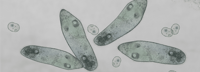
Microscopic image of paramecium and amoeba to represent live cell imaging techniques
Microscopy and Imaging | Lumenera

An Introduction to Live Cell Imaging

ByLumenera

Read on to learn more about live cell imaging, including how high rate microscopy can help you capture rapid cellular processes.

Read More An Introduction to Live Cell ImagingContinue

Man holding a lens up to the light to represent lenses and objectives
Microscopy and Imaging

How History Shaped Modern Optical Microscopes, Part Two: Corrected Lenses and Objectives

ByMartin Wilson

Discover how chromatic and geometric imaging aberrations have been corrected over the last few centuries with the development of corrected lenses and objectives.

Read More How History Shaped Modern Optical Microscopes, Part Two: Corrected Lenses and ObjectivesContinue

Vintage microscope to represnet the history of simple and compound microscopes
Microscopy and Imaging

How History Shaped Modern Optical Microscopes, Part One: Simple and Compound Microscopes

ByMartin Wilson

Discover the history of simple and compound microscopes in this first of our two-part series on the history of microscopes.

Read More How History Shaped Modern Optical Microscopes, Part One: Simple and Compound MicroscopesContinue

Image of a lumberjack cutting a tree to represent tissue sectioning tips
Microscopy and Imaging

Advanced Sectioning Techniques: How to Section Difficult Tissues

ByDr Jennifer Redig

Are you having problems with tissue sectioning? Follow these 10 tissue sectioning tips to create the perfect tissue section every time without stressing out.

Read More Advanced Sectioning Techniques: How to Section Difficult TissuesContinue

Image of vintage stereo as a play on words to represent a stereo microscope
Microscopy and Imaging

Stereo (Dissecting) Microscopes 101

BySteven Miller

Want to know about stereo microscopes? This article answers what a stereo microscope is, how it works and why it’s a great tool for biologists!

Read More Stereo (Dissecting) Microscopes 101Continue

multicoloured cells imaged on a confocal microscope to illustrate how you can improve your confocal images
Microscopy and Imaging

Confocal Images: Expert Tips for Your Hour of Need!

ByDr Nick Oswald

Getting publication perfect confocal images can be tricky. If you are struggling or just want to ensure you’re capturing the best images possible, check out our top 7 tips for confocal imaging.

Read More Confocal Images: Expert Tips for Your Hour of Need!Continue

light being defracted by a prism to represent optimizing the refractive index in confocal microscopy
Microscopy and Imaging

Resolution and Refractive Index: Set-up Your Confocal Wisely!

ByMelanie Laederich

Get the best out of your time on the microscope by understanding the refractive index of your experiment to optimize and increase your resolution.

Read More Resolution and Refractive Index: Set-up Your Confocal Wisely!Continue

Image of a iced coffee (Frappe) as a play on words for the microscopy technique FRAP
Microscopy and Imaging

Fun With FRAP! Fluorescence Recovery After Photobleaching for Confocal Microscopy

ByCatriona Paul

If you’re thinking FRAP is short for frappuccino then you need to read this article. Discover the history, how it works, and why you’d want it in your confocal toolbox

Read More Fun With FRAP! Fluorescence Recovery After Photobleaching for Confocal MicroscopyContinue

Colourful palms to represent Photoactivated Localization Microscopy (PALM).
Microscopy and Imaging | Zeiss

An introduction to Photoactivated Localization Microscopy (PALM)

ByZEISS Microscopy

How does photoactivated localization microscopy (PALM) work? And what use can PALM microscopy be to you? This short introduction to PALM gives you the answers!

Read More An introduction to Photoactivated Localization Microscopy (PALM)Continue

Page navigation

1 2 3 … 6 Next PageNext
Accelerate your research career with curated daily lab wisdom

We collate wisdom and tools from researchers worldwide to help you to accelerate your progress.

Sign up now to get it in your inbox

Webinars

Podcasts

Newsletters

Articles

Downloads

About Us
  • About Us
  • Meet the Mentors
  • Get Involved
  • Contact Us
  • Get the T-Shirt
Education
  • Technical Skills
  • More Skills
  • Events
  • Podcasts
Marketing
  • Life Science Marketing
  • Community Marketing
  • Custom Marketing
Bitesize Bio Powered
  • Microscopy Focus
  • Privacy Policy
  • Cookie Policy
  • Terms of Use

Copyright © Science Squared – all rights reserved

10 Things Every Molecular Biologist Should Know

The eBook with top tips from our Researcher community.

Get The Tips
Scroll to top