Choosing The Right Blood Collection Tubes
Selecting the right blood collection tubes for your experiment is crucial. But do you know what tubes to use for which type of blood sample? Read on to find the answers.
Join Us
Sign up for our feature-packed newsletter today to ensure you get the latest expert help and advice to level up your lab work.

Selecting the right blood collection tubes for your experiment is crucial. But do you know what tubes to use for which type of blood sample? Read on to find the answers.

Bioinformatics and NGS go together like peanut butter and jelly. But if you’re just starting out with these techniques it can be daunting.

During my first year of graduate school, I learned how to isolate bone marrow. I remember watching my mentor in awe, wondering how would I be able to do such a difficult technique. Flash forward to a few weeks later and I was confidently undertaking bone marrow isolation. Learning a new technique is always daunting,…

A Spot of History Most of the biomedical methods used started as a curiosity. Then the one-off gains a limited use, the technology then progresses until its use becomes widespread. Just think about the arch from the curious polished glass spheres, used by Antony Levnhook to look at animalcules, to modern microscopes. The same story…

If you look at the composition of peripheral blood, using hematology microscopy, you’ll see that it’s composed of multiple different cell types, including monocytes. It’s possible to isolate these different components to study and experiment on them directly. So, if you’ve done a few experiments and had fun with THP-1 cells, you can move on…
You may have heard about a breakthrough cancer therapy that engineers patient’s immune cells to fight their cancer using chimeric antigen receptor (CAR)-T cells. If you don’t live in the world of immunology, you may not know what a CAR is, or what it is used for. Here you’ll find a brief guide to CARs,…
I have worked in flow cytometry for a number of years. I’m still annoyed that many myths and imprecisions are perpetrated and perpetuated. Here is my non-exhaustive list of cytometry-related beliefs that send flow cytometrists screaming from the room or at least, being English, make me tut sadly. Forward Scatter Equals Cell Size No No…
In my previous article I discussed steps you can implement to ensure that a sample is ready for cell sorting. But now it’s time to make sure the sort worked. Here are a few sorting checks and measures to ensure that all’s well that ends well. Post-sorting Checks and Measures Re-evaluate Your Catch Tubes Sorting…
Flow cytometry is a pervasive tool to characterize just about anything in cell biology. From quantifying the expression of surface antigens, to determining the physiological changes in cells and everything in between, flow cytometry is as indispensable to a cell biologist as a knife is to a surgeon. Cell sorting is pivotal in enabling researchers…
As any good biologist knows, one of the easiest ways to determine if a cell is functionally active is the production and secretion of proteins in response to a stimulus. In many circumstances, the quantity of the secreted protein, and thus the level of cellular activation can be assessed by ELISA. However, if you are…

Hello again, fellow Flow Cytometry Fan! It looks like you have your experiment all planned out, including staining protocols and gating schemes, and are ready to get some paradigm-shifting data. But before we start “plugging-and-chugging” samples through your cytometer of choice, we need to make sure that the nozzle size and sheath pressure are set…

Flow cytometry is fast evolving from a method only revered by immunologists, to one used by nearly every biological specialty. It’s pretty much my favorite tool. Unfortunately, as with most lab techniques, much of flow cytometry is taught on the job without a lot of standards. And too often bad habits are passed along like…
Every bio- scientist who wants to analyze DNA knows that the process begins with the extraction of DNA from cells of interest. These cells could be RBCs, parasites, or bacteria to name a few. Furthermore, there are various DNA extraction methods1 to choose from depending on sample type, downstream analysis, and so forth. Many scientists…
Your reagents should do ‘Exactly what they say on the tin.’ This only happens though if you look after them in the way the manufacturer states on their data sheets. We have all been guilty of using reagents past their expiration date. Usually we can get away with it, but there are a few things…
Ah, cell counting — it’s the oldest trick in the book! Well, not really, but people have been developing methods for counting cells since the late 1800s. It has been around for a while. But what different methodologies are available to biologists now? Well, hold on, because you’re in for a treat! In this article, we…

There are several methods you can use to see if your T cells are cytotoxic, but a chromium release assay using radioactive 51chromium (51Cr) is one of the oldest. It gives good results, and is great for labs that can’t afford or don’t have flow cytometry readily available. Here, I will outline a simple method…
Multi-parameter data acquisition is key to the modern era of science research. I, for one, wish every single experiment that I design would give me the maximum amount of information. For example, in cell biology and immunology, we want to capture as much information (be it cytokines/hormones/chemokines) as possible about a given cell population. Of…
If you study human disease, you will likely handle a pre-analytical sample or two (or hundreds). For example, you could handle whole blood, serum or plasma, tissue biopsies, urine, fecal samples, cerebrospinal fluid, or synovial fluid—to name a few. You will probably use these samples to look for specific metabolites, proteins, or nucleic acids that provide…

To many users, the flow cytometer is a magic box: put in cells, get out data. You click the button to tell it which colors to look at without much thought about how the machine does this. However, not all fluorophores are created equal—some configurations might exclude the spectrum you’re really looking for. Here’s a…

While using human clinical samples in your research can provide robust and heterogeneous results applicable to larger portions of the population, working with these samples presents its own set of challenges. Here are some tricks I have learned to help isolate and grow your cells of interest while eliminating stromal, blood, or other undesired contaminants….

If you’ve been keeping up with our recent series of articles, welcome back! If not, you can catch up on how fluorescence works or what not to do with your flow experiment. In short, we have been discussing fluorescent labels and their role in flow cytometry. Today, I’ll round out our discussion by touching on…
Flow cytometry. Some people love it—most hate it—but all can agree that it is one of the most powerful analytical tools immunologists possess. Here’s a quick refresher: as the name suggests, flow cytometry measures the physical and chemical characteristics of cells. This is accomplished by fluorescently labeling cell surface markers/proteins using antibodies conjugated to fluorophores….

As science is becoming more interdisciplinary, the tools we use to answer questions are also crossing party lines. Case in point: flow cytometry. Once a tool only used by “real” immunologists, flow cytometry is fast becoming a method by which numerous questions can be answered, from the length of a cell’s telomeres, to the state…
Phosphorylation Equals Cell Signaling! How do cells communicate and respond to their environmental cues? This question has been on the hot list for scientists ever since the discovery of the cell. Cells use signaling cascades based on biochemical reactions to deliver or receive messages. How cool is that? The major secret of cell signaling was…

Selecting fluorophores can be a tricky business, but we’ve got you covered in this handy how-to guide.

While many scientists are methodical and precise, some of us like to live on the edge. Read a protocol all the way through? No thanks, I’ll take my chances and guess what concentration of HCl I should use. Label my tubes with the correct content? Puh-lease – it’s much more exciting deducing which is which…

So you want to work with mouse B cells? Primary murine B cells are a difficult, yet fascinating system to work with and can help deepen your understanding of an immunological system. You can study many things with primary B cells, including: These cells can be fickle to work with, but here are a few…

The MTS cell viability assay is one of the most important yet often daunting assays to perform for researchers in cancer biology, immunology, drug delivery pharmacy, etc. This chromogenic assay is extremely dependable for assessing the effects of a drug on different cell lines. However, it is only an easy assay to master if you…
There are some wonderful toys in the lab that enable us to open up a whole new world in science. One of those is a rather pricey and an incredibly sensitive laser-based apparatus capable of counting and sorting cells, detecting biomarkers, and engineering proteins: the flow cytometer. By propelling cells through the path of the…
It strikes fear into the hearts of new cytometrists. Compensation. More fights have started over the proper way to compensate at meetings than anything else. This article will strive to shed some light on the principles of compensation, and equip you with the tools necessary to achieve compensation mastery for your research experiments. Compensation is…
In a previous article, we went over the basic understanding of the inner workings of a flow cytometer. It’s important to grasp the types of measurements that are being made and, perhaps more importantly, what measurements are NOT being made. For simplicity’s sake, we’re going to frame this discussion in terms of a classical flow…
Flow Cytometry is a great way of seeing how many of your cells express a particular marker and how much of it is there. We do this by measuring fluorescence, but, as with all measuring systems, there will be signal that we are always trying to measure the above the noise. The signal that we…
Take a look at the dotplot below, are you happy with the way it’s presented? Do you think that you could recreate that experiment? If you were a reviewer, would you accept that figure? Sure, it’s flow plot, it shows 3 populations of which two are gated. Read many journals and you will see data…
One of the key characteristics of cytotoxic cells (i.e. CD8+ T cells, natural killer cells) is the presence of pre-formed cytoplasmic lysosomal granules. These structures house perforin and granzyme; two molecules that are essential for the lysis of target cells. Upon effector cell activation, granules are polarized toward the target cell and the contents are…

Have you ever wondered what a cell’s life is like? Do they constantly communicate to each other or do they just go on their own daily business? There is an easier way and it involves super advanced molecular “crayon” technology.
One of the much sought after question asked by many researchers worldwide is – “What is the gene expression profile of a single cell within a heterogenous pool of cells?” While mass cytometry is the current ‘hot’ methodology for single cell analysis, the good old flow cytometry can help us perform rapid analysis of single…
It’s happened to us all, you are ready to run your samples on the cytometer and you can’t see your cells on the screen. Here are a few tricks to troubleshooting this: Cytometer vs. computer connection The different types of cytometers will need different orders for switching on the cytometer and computer. Some are cytometer…
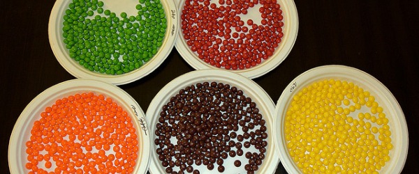
The flow cytometer that we have all grown to know and love may have only come into its own in the 1990’s, but who would have known that the first cell sorter was invented as early as the 1950’s? With the recent death of one of the key developers of fluorescence activated cell sorting (FACS),…

If you were to peek into a protein biochemist’s bag of tricks, what would you find? A mortar and pestle for collecting samples, some columns for isolating proteins and a mass spec instrument? Perhaps. But what about those little eppendorf tubes full of enzymes and helpful molecules? Certainly, each scientist has his/her own favorite. Here…
Part of my job in running a core flow cytometry facility is to make sure that the experiments that my users run have been optimised. But that optimisation can be split up into several areas. The first area is experimental planning: What do you want to know? Can you do this by flow cytometry? And…

The eBook with top tips from our Researcher community.