
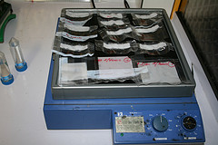
Subcellular Fractionation Protocols Explained
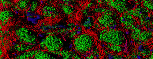
Perfect your subcellular fractionation in the lab by understanding how these protocols work. Plus, discover the various kits for organelle separation.
Join Us
Sign up for our feature-packed newsletter today to ensure you get the latest expert help and advice to level up your lab work.
Search below to delve into the Bitesize Bio archive. Here, you’ll find over two decades of the best articles, live events, podcasts, and resources, created by real experts and passionate mentors, to help you improve as a bioscientist. Whether you’re looking to learn something new or dig deep into a topic, you’ll find trustworthy, human-crafted content that’s ready to inspire and guide you.

Perfect your subcellular fractionation in the lab by understanding how these protocols work. Plus, discover the various kits for organelle separation.
Around and around the cell cycle goes, where it stops, nobody knows. Unless you have the right tools to analyze DNA content, that is. The DNA markers propidium iodide, Hoechst and DAPI are commonly used in flow cytometry to analyse a cell’s DNA content. Although they are simple to use, they do have disadvantages. Figure…

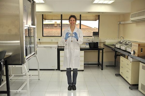
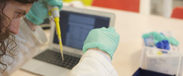
One of the most exciting aspects of being a biologist is getting opportunities to examine how and why living organisms behave the way they do. We have technology that enables us to obtain images at sub-cellular levels, and the skills to work directly with the micro-environments essential for the progression of life. However, at the…

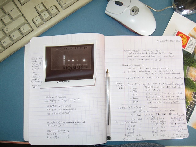
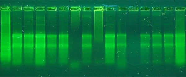
A wide variety of enzymes are available for PCR and RT-PCR and the optimal choice depends on a range of factors specific to your experiment. Some of these factors will now be explored to help you to make the most suitable and cost effective choices when ordering. PCR Type and Other Factors to Consider First,…
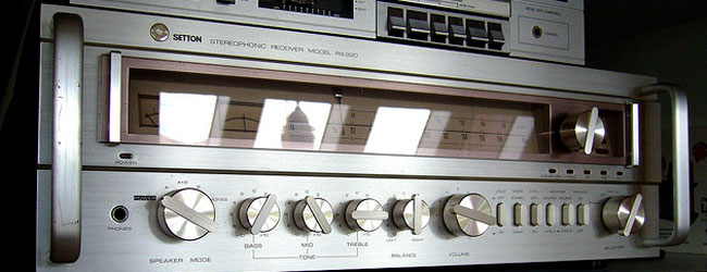
Ever tried to turn the volume all the way up on a small radio or small stereo system? (Hopefully you have not tried it with earphones in!) Notice how, after some point, the sound didn’t get any louder- it just got more distorted? That’s because you’ve hit the ceiling of your machine’s dynamic range. It’s…

What if there was a way to take the power and speed of a flow cytometer and couple it with the resolution of a microscope? Imaging flow cytometry does just that! Flow cytometry is a very powerful tool for the complex characterization of cells and cell populations but flow cytometers can also be thought of…

If you are struggling to optimise your Western blot protocol, one step to consider is the equilibration of your gel and membrane before transfer. Wondering what this step achieves and whether it’s necessary? You’re not alone! I did dozens of Westerns without ever bothering to equilibrate before I realised that it was having a big…

You are sitting in a seminar when you glimpse it: the figure that beats all other figures – a beautiful contour plot – and you realise that a similar figure is what you need to publish your paper in a high-ranking journal. You know the basics of flow cytometry, but admit you need a little…
So you’re designing a new experiment that requires PCR quantification. You used to have only one method to choose from, but now you have two – Quantitative Real-Time PCR (qPCR) and Digital PCR (dPCR). Which one is right for your application? Both methods have good quantification, sensitivity and specificity for most applications. They are compatible…
Stimulation of cells/tissue with a given stimulus (e.g., a cytokine) is a common experimental setup in any cell biology lab. The cellular response to the external stimulus e.g., the activation/deactivation of intracellular signaling pathways and/or the secretion of proteins is often the research goal, and there are a number of different methods that you can use to analyze such…

Bitesize Bio has had a lot to say about RNA isolation, mainly because it is one of the most anxiety-producing requirements for molecular biology; especially when you are first starting out (although isolating proteins from complex samples like soil and stool is far more difficult, let me tell you. But that’s a future post.) We’ve…
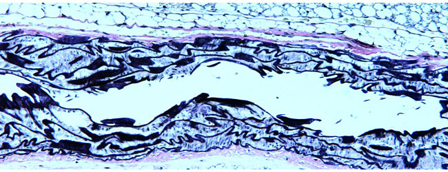
If you want to visualize elastic fibers in your sample, you need to use Verhoeff-van Gieson stain. Find out more about this stain, including how to use it.
You spent the last few weeks tweaking your Co-immunoprecipitation conditions, testing different antibody/bead combinations, and sampling a panaply of solutions and FINALLY! You have your Co-immunoprecipitation (Co-IP) elution… Now what? Well, you have a few choices. It really all depends on what you need know about the proteins in your elution. Do you need to identify…
The flow cytometer that we have all grown to know and love may have only come into its own in the 1990’s, but who would have known that the first cell sorter was invented as early as the 1950’s? With the recent death of one of the key developers of fluorescence activated cell sorting (FACS),…
Every PCR battle is the same: Too little amplification of your target DNA versus too much amplification of off-target DNA. But you can win the PCR battle and amaze your co-workers by mastering the use of PCR additives. PCR additives usually work one of two ways: 1) By reducing secondary DNA structures and thus increasing…
“A two photon microscope has higher sensitivity than a normal confocal microscope, because it uses two photos instead of one!” Yes, I can bear witness that this phrase has actually been uttered, and it was not by an undergraduate student. No exception to the rule The condensation of various levels of misunderstandings in this statement…
Ever had a blot so bad it looked like a Rorschach test? We have ten things that might be going wrong with your western blots and how to fix them.
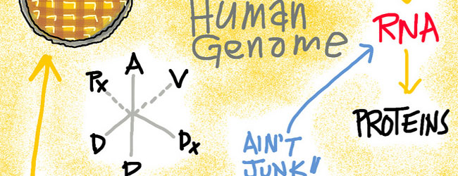
DNA sequencing (PCR, Sanger or next-generation sequencing (NGS)) is a now familiar part of any molecular biology lab. But ‘RNA-seq’, the so-called “Cinderella of genetics”, is now becoming the belle of the ball, providing new insights into this most central molecule of the ‘central dogma’. The many flavors of RNA Whilst genomic DNA is the…
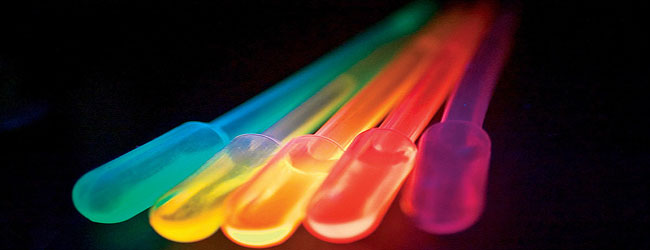
If you remember from one of my previous articles (if not, you can read it here!), we introduced ‘fluorophores’. These are basically substances (natural or synthetic) which have the ability to absorb light at a low wavelength and re-emit at a higher wavelength. In other words- they fluoresce! In this article, I’ll introduce the three…
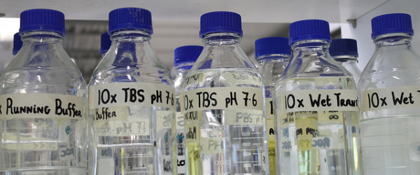
Buffers are often taken for granted, but they can make or break an experiment. In previous posts, we’ve talked about the wide ranges of buffers available for biological research and the characteristics of a “Good” buffer. Organic buffers are not inert! They can interact with your experimental molecule, or change pH due to changes in…
As widely used as it is, isolating RNA remains one of the more finicky protocols. Just about anyone who has performed the technique has their own personal tips and tricks to successfully isolate intact RNA from their samples with consistency. Although RNA can be somewhat unpredictable since it is so labile, there are a few…
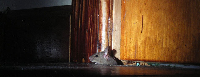
The issue of mouse-on-mouse background is only a cause for concern for the histotechnologist working within a research environment. Those working in a diagnostic setting will probably never experience this as they will be working with human tissue with antibodies raised in a variety of species- but one species that won’t be used is human!…

Do you wonder if your favorite protein interacts with another protein? Do you wish that you could shine a spotlight on your protein to determine its binding partner? You can use co-immunoprecipitation (Co-IP) to find your protein’s partner. This article will get you ready for your first Co-IP, provide a handy Co-IP protocol, and discuss…
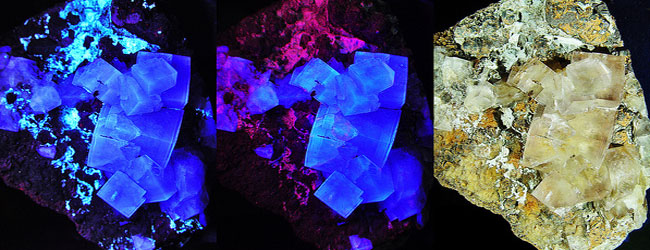
You may already use fluorescence as a tool in your microscopy and imaging work, but, do you know exactly what it is? Why are certain proteins and probes fluorescent? What causes this light emitting property? We’ll have a look at these and more questions in this article. Start with a definition We’ll start with a…

Western blots can be ugly. I mean down-right, horrifically, wall-of-shame ugly. Not only can they be embarrassing to show to your colleagues, but the ugliness can obscure your results, making it impossible to interpret your data. Blotting consists of many experimental steps, which makes the technique naturally error-prone. Although standardized protocols exist, many fail to…

Sometimes a clever little trick for cloning comes along that makes you just give an appreciatory “ahhh.” For me, it was TA cloning. TA cloning is not a new technique (I am showing my advanced laboratory age here), but when I discovered it, the simple elegance of the technique made me pause and wonder about…
Digital PCR (dPCR) is a quantitative PCR method that provides a sensitive and reproducible way of measuring the amount of DNA or RNA present in a sample. This method is similar to qPCR in the reaction assembly components and amplification reaction, but differs in the way the sample target is measured. Digital PCR is a…

Like most things in this world, fluorophores are mortal, and eventually your once bright fluorescent image will inevitably fade to black. This fading or ‘photobleaching’ of fluorescent signal can make imaging difficult, especially if you are trying to take quantitative images. Read below to learn what causes photobleaching of your fluorophores and how best to…
An ELISA (Enzyme-Linked ImmunoSorbant Assay) is a popular assay that uses antibodies and color change to detect proteins, peptides, antibodies or biomolecules in complex mixtures. ELISAs are popular because they are reliable, specific, easy to use, and can easily be scaled up to process multiple samples simultaneously. How an ELISA is Done: In an ELISA,…
We all know that stars are easier to see against the dark background of the night than they are to see against the bright sky of day. Well did you also know that the same may be true of your microscope specimen? Dark field microscopy is a popular microscope technique that makes your unstained transparent…
Every hard-core biologist knows designing the perfect construct can be a complex puzzle to solve. This challenge, if successful, can be extremely satisfying but can also drive you crazy for weeks. Luckily, Dr. Daniel Gibson and his colleagues at the J. Craig Venter Institute designed a new easy-to-use cloning method. Better yet, this system allows…

Although his name could fit in easily to the early 1980’s Hip-Hop Scene, Jerzy Nomarski (or ‘George’) was actually a Polish physicist with an interest in optical theory. Born in 1919, he eventually became a member of the Polish Resistance fighting in the Second World War. He was captured by enemy forces and held as…

While confocal microscopy seems to have become pervasive in cell biology, widefield microscopy techniques still have a special and important place. This month on the Microscopy and Imaging Channel, we’re focusing on widefield microscopy techniques: covering the basics of what these techniques are and when you should turn to them. What is this ‘Widefield’ you…

Bisulfite pyrosequencing is becoming a routine technique in molecular biology labs as a method to precisely measure DNA methylation levels right down to the single base. The technique allows for detailed and high resolution analysis of DNA methylation at specific genomic regions. How to detect the 5th base? Methylation of any of the four nucleotides…
You’re a senior grad student or postdoc, and you’ve done more PCRs than you can count. A new student has joined your lab, and you’ve been charged with training them on PCR. You don’t want to lead him/her astray, but it’s hard to remember the parts that you struggled with in your early days. This…
Phase contrast microscopy is a light microscopy technique which is primarily used to visualise live cells. Using various filters and condensers, the image produced by phase contrast allows us to see greater detail in live cells and can highlight aspects such as intracellular structures. Keep your cells alive! The best way to view cells is…

For those of us who work with Mus musculus or Homo sapiens, to name a couple of species, a few clicks on UCSC Genome Bioinformatics Site or Ensembl gets you the full and precise DNA sequence for any annotated gene in the genome. This luxury is not in place for all species however; many of…

Biologists have long appreciated the complexity of genome organization, but until recently lacked the tools to discern the intricacies of this puzzle. Now, thanks to some handy cross-linking, careful amplification, and (of course!) next generation sequencing, teams from Massachusetts are taking us down the rabbit hole, with some surprising findings from Wonderland. Bend Over Backwards…

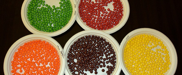
Discover the magic of toluidine blue – a polychromatic dye that changes color depending on which tissue component it is staining.
Blunt-end cloning involves the ligation of DNA fragments – usually between a plasmid vector and an insert – whose terminal ends are not “sticky”. Performing these ligations is notoriously difficult, particularly with large DNA fragments. But it is possible. And in this article I’ll give you some tips that I hope will increase your chances…

Technique, tips, ergonomics, and ISO guidance all in one handbook