4 Fixatives for Histology and Cytometry. Perfect Your Preservation
Learn about four fixatives for histology, which one you should pick, and how. Plus, get some top tips for perfect sample preservation.
Join Us
Sign up for our feature-packed newsletter today to ensure you get the latest expert help and advice to level up your lab work.

Learn about four fixatives for histology, which one you should pick, and how. Plus, get some top tips for perfect sample preservation.

Do you need to learn about gas chromatography? This article takes you through the basic principles and instrumentation. With illustrations!

With the NanoDrop spectrophotometer, quantifying a DNA, RNA, or protein sample concentration is as easy. Here’s everything you need to know about the strengths and limitations of this handy spec.

There are several great protein staining methods, but how do you pick the one that’s appropriate to your intended application? Read on to find out.

Flow cytometry is fast evolving from a method only revered by immunologists, to one used by nearly every biological specialty. It’s pretty much my favorite tool. Unfortunately, as with most lab techniques, much of flow cytometry is taught on the job without a lot of standards. And too often bad habits are passed along like…
Your reagents should do ‘Exactly what they say on the tin.’ This only happens though if you look after them in the way the manufacturer states on their data sheets. We have all been guilty of using reagents past their expiration date. Usually we can get away with it, but there are a few things…
In this article, you will be introduced to the world of fluorescent western blotting. Firstly, we will compare fluorescent and chemiluminescent western blotting. Then, we will learn how infrared fluorescent western blotting can give you truly quantitative and reproducible results. Lastly, we’ll look at the many advantages of fluorescent western blotting, including the possibility to multiplex. Importantly,…
You might have come across protein glycosylation before. Somewhere in the recesses of your memory you might even recall reading something about the protein you’re studying being glycosylated, but what does this mean and how do you analyze it? Glycosylated proteins are molecules decorated with sugar groups as they pass through the ER and Golgi…
Multi-parameter data acquisition is key to the modern era of science research. I, for one, wish every single experiment that I design would give me the maximum amount of information. For example, in cell biology and immunology, we want to capture as much information (be it cytokines/hormones/chemokines) as possible about a given cell population. Of…
While using human clinical samples in your research can provide robust and heterogeneous results applicable to larger portions of the population, working with these samples presents its own set of challenges. Here are some tricks I have learned to help isolate and grow your cells of interest while eliminating stromal, blood, or other undesired contaminants….

As science is becoming more interdisciplinary, the tools we use to answer questions are also crossing party lines. Case in point: flow cytometry. Once a tool only used by “real” immunologists, flow cytometry is fast becoming a method by which numerous questions can be answered, from the length of a cell’s telomeres, to the state…

Speed up your SDS-PAGE with our time-saving tips and tricks!

How do you pick which antibody you should use in your assay? If you’re starting a new assay and need an antibody for the job, then selecting a new antibody from the plethora available could be high up on your to-do list.
Every biochemist is familiar with proteases. More often than not, proteases cause a lot of anxiety. To this end, a lot of research has been done in developing techniques to prevent the activity of proteases. But some of these proteases can be the good guys too! For example, you can use them to separate your…
Sonication is mostly used during preparation of protein extracts to help break apart the cell. Although most lysis buffers have buckets of detergent that lyse cell membranes, sonication just gives an extra hand in breaking everything apart. Sonication also breaks up, or shears, DNA in a sample—preventing it from interfering with further sample preparation. Have…

Do you use biotinylated secondary antibodies in your immunohistochemistry? You could use polymers instead. They are a great time-saving reagent.
While you can observe mitotic cell cycle progression using immunofluorescence, flow cytometry is a great tool to delineate details that aren’t apparent by chromosomal morphology alone. DNA stains are a great way to get a general idea of what your cells are up to. There are also a number of other stains you can use…

Through many trials, and lots of error, I learned that there are many considerations for mass spectrometry that might not be obvious to you as a molecular biologist. Common contaminants, even in small quantities, can mask important peaks in your mass spec data and have a huge impact on the final results.
In this article I will not talk about ‘wild’ proteases, which destroy cellular proteins in your lysates like wolves destroy sheep. Instead, I’ll be talking about the shepherd dog proteases—purified, tame and useful to digest proteins your research. In Protein Research and Crystallization Several programs can predict your protein domains. However, we wet biologists know…
While many scientists are methodical and precise, some of us like to live on the edge. Read a protocol all the way through? No thanks, I’ll take my chances and guess what concentration of HCl I should use. Label my tubes with the correct content? Puh-lease – it’s much more exciting deducing which is which…

Fortunately for microscopy users, measuring intracellular fluorescence has been made relatively simple through an ImageJ plugin called the Cell Magic Wand. For those of you unfamiliar with ImageJ, it’s a popular image processing program that runs on Mac, Windows, and Linux. How to use ImageJ for measuring intracellular fluorescence First of all, to begin measuring…

Are you planning to do cellular immunology research? Then chances are you will be introduced to the flow cytometer – “a modern immunologist’s best friend.” This modern magic box is a highly versatile machine packed with cutting-edge fluidics and photonics (lasers). Combined with the monoclonal antibodies conjugated to fluorochromes capable of emitting light signals from a…

The whole TLC technique sounds easy to do, but it can be difficult and tricky during interpretation or give unexpected results, especially when working with biomolecules. For this reason, it is important to be familiar with troubleshooting thin layer chromatography. Some of the common problems faced during TLC and their solutions are listed below: Solvent…
Mass Cytometry is a relatively new technology which has recently featured in many high-impact journals. You may have read about instruments including the CyTOF, CyTOF2, and more recently, the Helios. With these instruments becoming more widespread, you might find yourself asking, what is mass cytometry, and what can it do for you? The Basis: Conventional…
To isolate your protein, you are going to need to tag it with something that will allow you to fish it out of the bacterial protein soup. This is where transition metals can help you out!

What do you use if antibodies are too large for super resolution microscopy? Aptamers. These are small affinity reagents (~ 2 nm!) that interact with their target in the same way as an antibody, but without the hefty backbone attached.
Most eukaryotic proteins exist as several isoforms, differing in posttranslational modifications, which allows them to perform slightly different functions or the same function under slightly different conditions. A common posttranslational modification of proteins is glycosylation.
Formalin fixed paraffin embedded (FFPE) tissues are valuable samples that typically come from human specimens collected for examination of the histology of biopsies for the detection of cancer. But each sample contains much more information just waiting to be unlocked. Despite the tiny sample size, DNA can be extracted from the tissue sections and used…
We’re already gone through the basics of how gel electrophoresis work, compared common gel types like agarose and polyacrylamide and even explored some alternatives. Now let’s look at the native versus denaturing gels. You’ll be a speGEList in no time! Denaturing Gels We’ll start with this one, as it’s very self-explanatory. Denaturing gels are exactly…

First of all let me say the technique of labeling tissues (immunohistochemistry, IHC), and cells (immunocytochemistry, ICC) is indeed immunoscience NOT alchemy, though at times it may certainly seem like alchemy! But to scientists inexperienced in this technique, who typically see the results of IHC/ICC experiments in the form of pretty pictures, it can certainly…
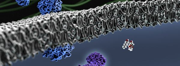
Supported lipid bilayers are a very useful tool in many fields of cell and membrane biology. But how easy is it to make them? Bilayers can be made quite reproducibly, once you have found a reliable protocol! However, it can take some time to optimize your technique, so to increase your chances, make sure you…

Like most other chromatographic techniques, thin layer chromatography (TLC) separates out individual compounds from a mixture depending upon the polarity of each compound. The solvent system travels up a silica plate by capillary action and passes over the sample that you spot onto the plate. As the solvent travels up, it moves the compounds present…
While working as biologists, we often come across mixtures of compounds, and the first question that strikes our minds is ‘what are the components in this mixture?’ One might think of using chemical assays to find the presence of specific compounds. But that sounds painful, doesn’t it? Well, the good news is that thin layer…

As biochemists, we routinely run SDS-PAGE to analyze our proteins. Imagine the time and effort you are going to save when you can run every gel to perfection.

While most of us have heard of super resolution microscopy, many of you may not have heard of MSIM, or Multifocal Structured Illumination Microscopy. This under-the-radar imaging technique is relatively quick, cheap (by comparison) and will allow you to get a lot of data, fast. So What is MSIM Anyway? MSIM, as I mentioned earlier,…
Are you an assiduous biologist who prefers label-free imaging methods for biological samples analysis? Raman spectroscopy offers you a wonderland of imaging technique with unlimited benefits. To start with, Raman Spectroscopy is a spectroscopic technique based on inelastic scattering of monochromatic light usually from a laser in the visible or near infra-red part of electromagnetic…

Western blotting uses electrophoresis and antibody-epitope affinity to give a semi-quantitative and (theoretically) clear measure of protein abundance. It’s a long procedure, filled with many steps—and even more room for error. Learning to troubleshoot certain problems is incredibly important for continued success with this technique. So what do you do when your final imaged product…
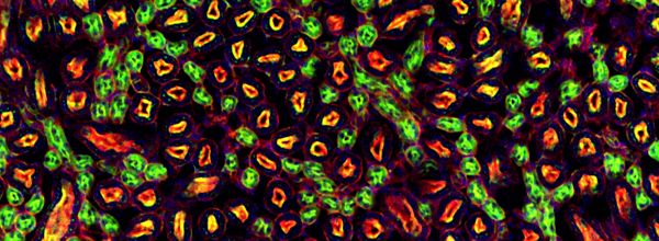
Taking publication quality immunofluorescent images of can be a very time intensive, and frustrating process with hours spent capturing, processing, and putting the images into final figure format. And, if you aren’t careful, you can do a lot of work only to realize later that you need to re-image something for one reason or another….
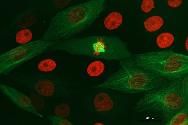
Even in the most basic applications, fluorescence microscopy can be a very powerful technique. Simply put, the ability to actually see the biology you are interested in cannot be matched in directness. Often, the aim of fluorescence microscopy is to observe the effect of an experimental manipulation. Ultimately, you would like to know that the…

Want to save money on one of the most expensive steps of your Western blots? Then, use less antibody for significant cost savings!

The eBook with top tips from our Researcher community.