While most of us have heard of super resolution microscopy, many of you may not have heard of MSIM, or Multifocal Structured Illumination Microscopy. This under-the-radar imaging technique is relatively quick, cheap (by comparison) and will allow you to get a lot of data, fast.
So What is MSIM Anyway?
MSIM, as I mentioned earlier, stands for Multifocal Structured Illumination Microscopy. This means that the instrument uses more than one focal point of light, in a structured pattern, when it images. It is also a super-resolution technique, meaning you can image below the diffraction limit of light. It is the little sister of SIM, but you won’t see if for sale through Zeiss or Olympus, as they are generally adapted from other microscope systems.
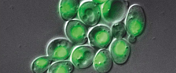
MSIM has an astounding resolution of 145nm in the transverse direction and 350nm in the axial direction. This means you can make out some of the really small structures inside cells, such as organelle membranes and cytoskeleton structures. It can even be used to image relatively thick samples of about 50um!
How Does it Work?
MSIM relies on a scanning, structured set of light points. Think of it as a grid of focal points that passes over where you are imaging. This allows the instrument to essentially add more “points” and collect more data while it takes the image allowing you to get as much data from your field of interest as possible. The light pattern also helps to reduce out of focus light, allowing for greater resolution.
The drawback to MSIM is that the raw data is virtually unreadable. The data generated by this method requires digital post processing. The post processing uses Fourier transforms to determine where the fluorescence is centered and convert the gibberish raw data into a nice pretty image you can actually use and analyze.
Why MSIM?
MSIM is a relatively cheap method of super resolution Imaging. For a lab on a budget (which I think is most of us!) you can save money by adapting an already existing microscope and building an MSIM yourself with easily orderable parts. While a specific list of parts is not readily available, the Shroff lab at the NIH has all the know-how you need.
The MSIM is also incredibly fast, imaging over 100 2D slices per second. This method allows you to generate a lot of data, very quickly. So if you are limited on time and money, but want great quality images, then MSIM might be a good fit for you.
If you are interested in learning more about this technique, you can check out this paper on MISM published in Nature.
What are your thoughts on MSIM? Would you use it? Have you used it? Comment below!


