How to Become a Live Cell Paparazzo: A Beginner’s Guide
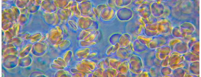
Think of the very first time you looked at cells under a table-top microscope. Here’s what you would have done in that experiment: Step 1: Grow cells. Step 2: Plate cells on to a glass/quartz slide. Step 3: Insert the slide under a microscope and look. The protocol for performing single-cell microscopy has a similar…