Tissue Processing For Histology: What Exactly Happens?
Tissue processing for histology is a key step between fixation and embedding. We take you through the steps of tissue processing in this simple guide.
Join Us
Sign up for our feature-packed newsletter today to ensure you get the latest expert help and advice to level up your lab work.

Tissue processing for histology is a key step between fixation and embedding. We take you through the steps of tissue processing in this simple guide.

Oil immersion microscopy can improve your resolution in microscopy. This article will explain why this is the case and how you can use oil immersion microscopy in the lab!

Discover the history of histology, from the first mention of a cell in 1665 to the identification and development of various stains.

There are a large number of microscope objective abbreviations relating to optical aberrations; here we’ll shed some light on some of the most common ones to get you up to speed in no time!

Discover how chromatic and geometric imaging aberrations have been corrected over the last few centuries with the development of corrected lenses and objectives.

Discover the history of simple and compound microscopes in this first of our two-part series on the history of microscopes.

Have you ever been looking through a box of slides and found something that you want to image or look at later, or even show to one of your colleagues or supervisor? Finding that exact spot on the slide at a later date can prove to be difficult- using a marker pen on the coverslip…
Keeping up to date with the scientific literature is a large part of the work-load of any researcher. Love it or loathe it, this means of sharing research findings with the larger scientific community is still the way in which most of us inform ourselves of the latest findings in our fields of research, or…
Photography has undergone great improvements in the last few decades. In times gone past, photographic film was used. Now most researchers use digital means to capture their images. But not all digital cameras are the same. For optimal results you need to know the different types of microscope cameras and how they work. Before the…
How often have you looked at slides down the microscope and your thoughts have been miles away? Have you ever been sitting at the bench pipetting and preparing a PCR and wondered if you had really added your forward primer to all your samples (I’ll put my hand up to this one!)? Or spent time…

When you first start out using a microscope, you might only adjust the eye pieces, objectives, and the focus controls. However, you shouldn’t overlook the microscope condensers as they are an important part of the whole optical system of a microscope.
If you want to make molecules stick together you need to know about streptavidin/biotin. This article follows on from Mike’s article looking at ‘sandwich’ and ‘amplification’ methods of immunohistochemistry (IHC) and covers how streptavidin-biotin works in IHC, including protocols. Streptavidin-Biotin What is it? Avidin is a natural biotin-binding protein found in egg whites. Streptavidin is similar…

When you fix your tissue samples with paraformaldehyde (PFA) the proteins in your sample become covalently cross-linked. This is good to preserve the ‘architecture’ of your tissue sample. However, this cross-linking can become a problem when you carry out immunohistochemistry (IHC). Cross-linking can ‘mask’ or hide your antigens-of-interest and make them ‘invisible’ to your IHC…

The magnification and viewing of samples using a microscope relies on both the objectives and the eyepieces working harmoniously together. If you buy a ready-to-use microscope, then the objectives and the eyepieces which are fitted as standard will be designed to complement each other. On the other hand, if you are designing and building a…
Two different sensors are generally used in cameras for microscopy: Charge Coupled Devices (CCD) or Complementary Metal Oxide Semiconductors (CMOS or sCMOS). Although there are a number of similarities between the two sensors, differences in the way they function can have an effect on image capture time as well as signal-to-noise ratio. Let’s take a…
If you remember from one of my previous articles (if not, you can read it here!), we introduced ‘fluorophores’. These are basically substances (natural or synthetic) which have the ability to absorb light at a low wavelength and re-emit at a higher wavelength. In other words- they fluoresce! In this article, I’ll introduce the three…
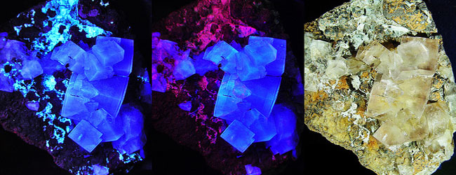
You may already use fluorescence as a tool in your microscopy and imaging work, but, do you know exactly what it is? Why are certain proteins and probes fluorescent? What causes this light emitting property? We’ll have a look at these and more questions in this article. Start with a definition We’ll start with a…

Although his name could fit in easily to the early 1980’s Hip-Hop Scene, Jerzy Nomarski (or ‘George’) was actually a Polish physicist with an interest in optical theory. Born in 1919, he eventually became a member of the Polish Resistance fighting in the Second World War. He was captured by enemy forces and held as…
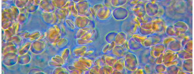
Phase contrast microscopy is a light microscopy technique which is primarily used to visualise live cells. Using various filters and condensers, the image produced by phase contrast allows us to see greater detail in live cells and can highlight aspects such as intracellular structures. Keep your cells alive! The best way to view cells is…

Most of the microscopes you will encounter in your laboratories will be ‘upright’. In other words, they are assembled (from top to bottom) in the order of; eyepieces, objectives (on revolving nosepiece), stage, sub-stage condenser, diaphragm and base. However, there are two other types of light microscopes you will perhaps encounter (and use) and it…

Following on from the first part of the H and E 101 articles, here are the materials and recipes you’ll need for your own H and E workstation (assuming you don’t have access to a histology lab). Many of the chemicals listed below are toxic and/or harmful. Use PPE when handling/storing, follow SOP’s in your…

Haematoxylin and Eosin staining is the most common staining in the modern (and old!) histology lab. This staining technique gives an overview of the structure of the tissue and can be used in pathological diagnosis. This article follows on from Nicola’s introduction, but we’ll take an in-depth look at the stains, chemistry and method to…

The theory behind the idea of having shared microscopes is a good one, but, in reality, this can sometimes mean you have to put up with the dirty habits of your fellow scientists and researchers. And some of your lab mates turn out to be really mucky! Here’s my Top 10 of things which really…
Following on from our previous article, here are some suggestions for an old microscope (should you happen not to destroy it!). 1. Museum piece Start your own mini scientific instruments museum. Before you know it, you be raking through the old skips and dumpsters at your institute looking for exhibits. 2. Teach kids Teach your…

You’ve spent days, perhaps weeks or months squirrelling away tubes of preserved tissue in the dark drawers under your laboratory bench like the trophies of a demented serial-killer. Hours have been spent in histology in the processing, embedding and sectioning onto slides. Finally, like a warrior victorious in battle, you hold aloft your thin glass…

The eBook with top tips from our Researcher community.