Traditionally, if you’re hoping to clone a DNA/RNA fragment (or insert) into a vector, such as a BAC you would need:
- Expensive exonucleases, called restriction enzymes: pacman-like enzymes that chomp at specific sequences in your destination vector or fragments to be inserted (often just “inserts”).
- Sequence homologies between your inserts and your destination vector, called restriction sites.
- A ligase: an enzyme that glues your sequence back together by filling in the gaps left once you’re insert has been incorporated into your vector.
This process looked something like this: cut you vector using exonucleases to expose ends that are complementary to the ends of your insert and then stick your insert into the vector using a ligase. This was an expensive and problematic process that has since reduced in popularity with the dawn of ligase-independent and/or restriction enzyme-free cloning methods like SLIC, CPEC, TOPO and Gibson (Gibson uses Taq ligase.)
For those who still cling on to their restriction enzymes and ligase singing that such ligase-independent methods can lead to small errors when you go to PCR to amplify your product, I give you proofreading enzymes like Phusion that will minimize this risk.
Introducing SLiCE
SLiCE is another ligase and restriction enzyme-free cloning method and is very similar the other methods mentioned above. First described by Zhang et. al in 2012, SLiCE uses bacterial cell extracts to assembly multiple DNA fragments into recombinant DNA molecules and all in the same in vitro recombination reaction. The trick: Short end homologies of 15–52 bp that can be used in the absence of or in conjunction with flanking heterologous sequences. This allows for directional subcloning of DNA inserts or fragments from BACs and other vectors.
Since many of the standard lab bacterial strains that you have floating around can be harvested to create your SLiCE extract, this method is a lot more cost effective than tradition cloning. Some strains are more efficient that others, for example PPY, a strain of E. coli DH10B that expresses a ? red recombination system. So why use this method? Let’s take a peak!
Reasons to Use This Method
- Cost effective for high throughput screening
- It’s “seamless”, meaning it doesn’t require you to add sites for enzymes to target and cut (called junction sites) so you don’t have unwanted sequences dotted around your product.
- It doesn’t require the use of expensive restriction enzymes or ligases.
- You can use fragments generated either by PCR amplification or restriction digestion.
- You can combine several steps into one as SLiCE allowing for the assembly of multiple DNA fragments in one cloning step. This makes it a great option for the assembly of multiple DNA fragments during gene synthesis experiments.
Sounds good to me!
What Are the Steps?
The main players here are your plasmid and your SLiCE extract.
Step 1: Design and create your plasmid.
Step 2: Select your E. coli strain and harvest cells from a stable, competent stock solution. Lyse these cells in lysis buffer and centrifuge to isolate the cell debris from the cloning insert DNA. Keep the supernatant and discard the pellet. These cell extracts can be stored in 50% glycerol at –80oC if necessary.
Step 3: Linearize the destination plasmid vector by PCR amplification or restriction digest and amplify the cloning insert of your interest using specially designed primers. These primers must contain 5’-ends homologous to the vector or to other cloning inserts (to create a string of attached fragments, although the ends of these must contain the 5’ homologous ends.)
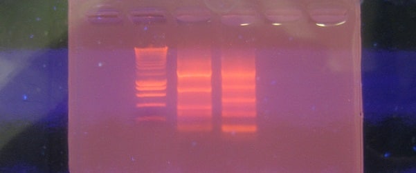
Step 4: Once the residual plasmid template DNA has been removed, the linearized vectors and PCR fragments can be tested for confirmation of successful generation using gel electrophoresis.
Step 5: Release your vectors and fragments from the gel using by purifying extraction.
Step 6: Incubate your vector and fragments in a solution of SLiCE buffer containing magnesium chloride, ATP, DTT and Tri-HCl with water at 37oC for 1 h.
Step 7: Transform your fragment-vector hybrids into competent cells with by electroporation or by chemical transformation.
Step 8: Isolate all cells that have successfully taken up the fragment-vector hybrids by plating in the presence of appropriate antibiotics or however you designed your vectors to be detected.
For more details, see the original paper and for a look into some of the restrictions and how to get around them.
This is a fantastic and simple method that shouldn’t take too much optimizing – yay for less moaning and more cloning!


