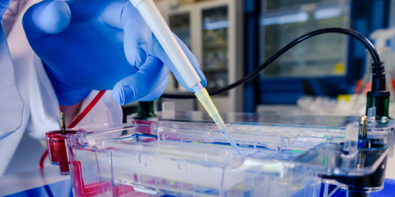
Reverse transcription optimization is one of the most overlooked aspects of RT-PCR best practices. The RT step converts RNA to cDNA, and it is one of the largest and most easily underestimated sources of technical variability in RT-qPCR.
The problem is that RT efficiency is never 100%. It varies across transcripts and is sensitive to conditions that most protocols treat as fixed. Two RNA samples with identical concentrations and purity ratios can produce cDNA of very different quality depending on primer choice, reaction setup, and template condition.
This article covers the six factors you can control, with the specific conditions and thresholds that matter for each one. If your reverse transcription is already failing, start with our article on common RT problems instead. If you need to decide between one-step and two-step workflows first, our one-step vs two-step RT-PCR decision guide covers that.
Before You Start: RT Setup Checklist
These are the conditions experienced researchers verify before every reverse transcription reaction. Skipping any one of them introduces compounding variability.
Choose a free resource to help you move forward
REFERENCE CARD
qPCR Efficiency & Ct Reference Card
EBOOK
The Fundamentals of qPCR and RT-qPCR
Reverse transcription setup checklist
- RNA purity confirmed: 260/280 between 1.8–2.1 and 260/230 above 1.8. If the 260/230 is below 1.5, re-purify before proceeding. Guanidine or phenol carryover will inhibit the reverse transcriptase.
- RNA integrity checked: RIN ≥7 is a safe routine acceptance criterion for RT-qPCR (RIN ~5 may be workable for short amplicons if all samples are similarly degraded). Alternatively, check for intact 28S/18S bands on a gel. Degraded RNA produces truncated cDNA and systematically under-represents longer or 5′-distal transcripts.
- RNA input standardized: Use the same total RNA mass (typically 0.5–2 µg) across all samples in the experiment. Varying input changes RT efficiency and makes between-sample comparisons unreliable.
- Priming strategy matched to your targets: Oligo-dT for eukaryotic mRNA with intact poly(A) tails. Random hexamers for degraded RNA, non-polyadenylated RNA, or prokaryotic targets. A blend for general-purpose eukaryotic gene expression.
- gDNA removal confirmed: DNase treatment completed (on-column or in-solution), or intron-spanning primers validated for every target. Include a no-RT control on every plate.
- One method for the whole study: Once you choose a priming strategy, enzyme, and workflow (one-step or two-step), do not change mid-experiment. RT efficiency differences between methods make cross-method comparisons invalid for quantitative work.
Factor 1: Choice of RT Primer
The primer you use for reverse transcription determines which RNA molecules get converted to cDNA, how completely they are transcribed, and how much of your enzyme activity is directed at your targets of interest. There are several options, each with a clear use case:
| Oligo-dT (12–18mer) | Eukaryotic mRNA with intact poly(A) tails | Misses non-polyadenylated RNA; 3′ bias with long transcripts (>4 kb); fails with degraded RNA | Standard eukaryotic gene expression |
| Random hexamers | Degraded RNA, prokaryotic mRNA, non-polyadenylated targets, 5′ regions of long genes | cDNAs are not full-length; primes rRNA and tRNA (reducing mRNA-specific yield); higher background | Prokaryotic work, FFPE samples, targets near the 5′ end |
| Gene-specific primers | Maximum sensitivity for a single target; one-step RT-PCR | One cDNA per primer; cannot re-use the cDNA for other genes | One-step RT-qPCR, low-copy detection, clinical diagnostics |
| Oligo-dT + random hexamer blend | General-purpose eukaryotic qPCR; balances 3′ coverage with internal priming | Slightly lower per-target yield than gene-specific priming | Multi-gene eukaryotic studies; recommended default for two-step RT-qPCR |
Which Primer should I use?
For most eukaryotic RT-qPCR experiments, a blend of oligo-dT and random hexamers gives the best balance of transcript coverage and representation. This approach captures both full-length poly(A) transcripts and internal regions that oligo-dT alone might miss on longer mRNAs. Several commercial RT kits include optimized blends for this purpose.
If your transcript is long (>4 kb), oligo-dT priming alone will under-represent the 5′ end. Random hexamers solve this, but the cDNA fragments will be shorter. Using longer random primers (8–9mers instead of 6mers) reduces priming frequency and increases average cDNA fragment length, which can help when your qPCR amplicon is positioned far from the 3′ end.
Note: The primer you use for reverse transcription determines which fraction of your transcriptome gets represented in the cDNA pool. Choose the wrong primer for your target and your qPCR will measure an incomplete copy of the real biology! So choose wisely.

Factor 2: RNA Secondary Structure
RNA folds into secondary structures (e.g., hairpins, stem-loops, and pseudoknots) that can stall or displace the reverse transcriptase enzyme during cDNA synthesis. When the enzyme hits a stable structure, it either falls off the template or pauses long enough to produce a truncated cDNA. The result is incomplete reverse transcription, which leads to lower apparent expression of that gene.
GC-rich regions are the strongest predictors of secondary-structure problems. If your target gene has a GC content above 60%, expect the RT step to be more difficult. You have two strategies for dealing with this:
- Thermal denaturation before RT. A 5-minute incubation at 65°C with the RNA and primers (before adding the enzyme) relaxes most secondary structures. Where compatible with your enzyme and kit protocol, this should be part of every RT setup. Snap-cool on ice immediately after denaturation to prevent structures from reforming before the enzyme binds.
- Thermostable reverse transcriptases. Enzymes that operate at higher temperatures (50–55°C instead of 37–42°C) can read through structures that would stall a standard MMLV-based RT. Current-generation engineered RTs — including thermostable variants from most major vendors — handle GC-rich templates significantly better than the older enzymes. If you routinely work with GC-rich genes, a thermostable RT is worth the switch.
The practical test: if you see consistently lower yields or later Ct values from one gene compared to expectations based on its known expression level, and that gene has high GC content, secondary structure in the RT step is the most likely cause.

Factor 3: Removal of Genomic DNA
Genomic DNA contamination in your RNA extract can lead to false positives. The gDNA co-amplifies with cDNA during the PCR step, inflating the apparent expression of your target. This is one of the most common sources of error in RT-qPCR, and MIQE/MIQE 2.0 reporting guidance lists gDNA removal as a required reporting element.
There are two complementary approaches, and the strongest strategy uses both:
- DNase treatment. Treat the RNA with DNase I either during extraction (on-column) or after extraction (in-solution with a heat-inactivation step or a cleanup column). On-column DNase is convenient but may not remove all gDNA from high-input samples. In-solution treatment can be more thorough, especially for high-input or gDNA-rich samples, but requires careful inactivation or cleanup to avoid DNase carryover into the RT.
The key point: do not skip DNase treatment for quantitative work. Even column-based RNA extraction kits that claim to exclude gDNA can still leave sufficient contamination to yield a signal in a sensitive qPCR assay. - Intron-spanning primers. Designing your qPCR primers to span an exon-exon junction means that genomic DNA (which contains the intron) either cannot be amplified or produces a product of a different size that can be distinguished on a gel. This is the most reliable way to exclude gDNA signal from your qPCR results for eukaryotic targets.
One important exception: pseudogenes. A pseudogene is a processed copy of a spliced mRNA that has been reverse-transcribed and reinserted into the genome. Pseudogenes lack introns, so primers designed to span intron-exon boundaries will still amplify them from gDNA. If your target gene has known pseudogenes, you also need DNase treatment. GAPDH, beta-actin, and many commonly used reference genes have pseudogenes.
For prokaryotic RNA, which has no introns, intron-spanning primers are not an option. Genomic DNA removal by enzymatic DNase treatment is the only approach, and it becomes critical for accurate gene expression measurements. A no-RT control must be included on every plate to confirm the DNase treatment was effective.

Factor 4: RNA Integrity
RNA quality has a direct, measurable impact on cDNA synthesis efficiency. Degraded RNA produces truncated cDNAs, under-represents longer transcripts, and introduces batch-to-batch variation that no normalization strategy can fully correct. Checking RNA integrity before reverse transcription should be routine for quantitative work.
How to check RNA Integrity
- Agarose gel: Intact mammalian total RNA shows sharp 28S and 18S ribosomal RNA bands on an agarose gel, with the 28S band approximately twice as intense as the 18S band (the theoretical ratio is ~2.7:1, but 2:1 is the widely used practical benchmark). Equal intensity between the two bands indicates mild degradation, but the material is often still usable. Heavy smearing, particularly toward the bottom of the lane, indicates significant degradation and RNA that will produce unreliable cDNA.
- Bioanalyzer: For a quantitative assessment, the Agilent Bioanalyzer or TapeStation provides an RNA Integrity Number (RIN) on a scale of 1–10. For short-amplicon RT-qPCR (<150 bp), a RIN around 5 may be workable if all samples are similarly degraded, but RIN ≥7 is a safer routine acceptance criterion. For longer amplicons or when cross-sample consistency matters, aim for RIN ≥8 and match degradation levels across all samples. The RNA quality for qPCR article covers purity thresholds, RIN interpretation, and assessment method selection in full detail.
If you do not have access to a Bioanalyzer, running 1–2 µg of total RNA on a 1% agarose gel provides a qualitative integrity check that is sufficient for routine work. It uses more samples, but it costs almost nothing and catches the gross degradation that would ruin your RT.
Factor 5: Accurate RNA Quantitation
Accurate measurement of RNA yield matters because you need to load the same mass of RNA into every reverse transcription reaction across your experiment. If one sample has twice the RNA input of another, RT efficiency will differ, and your downstream qPCR will reflect the input variation rather than true differences in expression.
How to quantify RNA
- UV spectrophotometry: Using a NanoDrop or similar microvolume instrument is the standard method. It requires only 1–2 µL, gives instant results, and measures concentration from absorbance at 260 nm. The limitation is that UV absorbance does not distinguish RNA from DNA, and genomic DNA contamination can inflate the apparent RNA concentration. Salt and phenol carryover from extraction can also absorb at 260 nm, giving falsely high readings.
- Fluorescent assays: If you suspect DNA contamination is affecting your concentration measurements, RNA-binding fluorescent assays, such as RiboGreen, are more sensitive than absorbance-based methods and are useful after DNase treatment. However, they are not completely RNA-specific, and residual DNA can still inflate measurements. For the most accurate quantitation, perform DNase treatment before measuring with either UV or fluorescent methods.
Whichever method you use, the important thing is consistency: use the same quantitation method across all samples in your experiment, and make the measurement after any cleanup or DNase treatment steps, not before.
Factor 6: One-Step or Two-Step RT-PCR
Whether you run reverse transcription and PCR in a single tube (one-step) or as separate reactions (two-step) affects sensitivity, flexibility, and the way you use the resulting cDNA:
- One-step RT-PCR eliminates the need for sample transfer, reducing contamination risk, and uses gene-specific primers for maximum sensitivity. It is the standard for clinical diagnostics and high-throughput screening, in which you measure one target per reaction.
- Two-step RT-PCR converts RNA to a cDNA pool that can be aliquoted across many reactions, allowing you to measure multiple genes from the same reverse transcription. This eliminates RT efficiency as a variable when comparing genes within a sample.
Once you choose a method for a study, stay with it. RT efficiency varies across one-step and two-step workflows, priming strategies, and enzyme formulations. Mixing methods within a quantitative study makes the data impossible to compare reliably. Our one-step vs two-step RT-PCR guide covers the full comparison, including when each method is the better choice.
What the Protocol Doesn’t Tell You
- The order you add components to the RT reaction matters more than most protocols suggest. Your RNA and primers should be combined first and heat-denatured together (65°C, 5 min, then ice). Only then should you add the enzyme mix, buffer, and dNTPs. If you add everything at once and heat the whole reaction, the enzyme is exposed to denaturing temperatures. Some enzyme formulations tolerate this, while others lose significant activity. The safest approach is the two-step addition: denature RNA+primers, then add the enzyme mix at the reaction temperature.
- Your RT enzyme is probably fine, but your RNA input consistency is probably not. When RT-qPCR results vary between replicates, the instinct is to blame reverse transcriptase. In practice, the most common cause is inconsistent RNA input, i.e., pipetting 1.8 µg into one reaction and 2.3 µg into the next, or using a NanoDrop reading that was inflated by gDNA. If you tighten your quantitation and standardize input, most “enzyme variability” disappears.
- Leftover cDNA degrades faster than you think at −20°C. Protocols say cDNA is stable at −20°C, but, in practice, repeated freeze–thaw cycles can introduce avoidable variability, especially for low-abundance targets. This can cause your Ct values to drift upward in ways that are difficult to distinguish from real changes in expression. Aliquoting your cDNA immediately after synthesis into single-use volumes prevents freeze-thaw. If you are running a large study over weeks, this one step eliminates an entire category of unexplained replicate variation.
- No-RT controls that are “clean” at Ct 35+ are not necessarily clean. A no-RT control with a Ct of 36 seems negative, but if your target gene Ct is 30, that is only a 6-cycle difference; assuming near-100% PCR efficiency, the gDNA signal contributes roughly 1.5% of your total signal. For most gene expression studies, this is acceptable, but for targets with Ct values above 32, even a “negative” no-RT at Ct 37 can represent significant gDNA contamination relative to the true signal. Your no-RT Ct must be at least 5–7 cycles above your target Ct to be considered negligible.
- Random hexamers prime ribosomal RNA (a lot of it!) When you use random hexamers, the vast majority of your cDNA is rRNA-derived because rRNA makes up 80–85% of total RNA. Only a small fraction of the RT enzyme activity goes to your mRNA targets. Random priming can give different Ct values from oligo-dT priming (often later) because much of the cDNA pool derives from abundant rRNA. This is expected behavior, but if you switch priming strategies mid-project, the Ct shift will look like an expression change. Do not compare Ct values across priming strategies without validation.
Common Mistakes
| Varying RNA input between samples | Replicate Ct values are inconsistent; reference gene Ct varies by >0.5 cycles across samples | Measure RNA concentration after cleanup; pipette from a diluted working stock at a fixed concentration |
| Skipping the 65°C denaturation step | Consistently lower yields from GC-rich targets; truncated cDNA products on a gel | Where compatible with your enzyme/kit protocol, heat RNA + primers at 65°C for 5 min and snap-cool on ice before adding enzyme |
| Using oligo-dT for degraded or prokaryotic RNA | No or very late Ct for targets known to be expressed; 3′-biased amplification pattern | Use random hexamers or a blend for degraded, prokaryotic, or non-polyadenylated targets |
| Omitting the no-RT control | Cannot distinguish cDNA signal from gDNA background; inflated expression for intronless genes | Include a no-RT control for every RNA sample on every plate; compare Ct to +RT samples |
| Relying on intron-spanning primers without checking for pseudogenes | No-RT control shows amplification despite intron-spanning primer design | BLAST your primer sequences against the genome; check for processed pseudogenes of your target |
| Switching RT method or enzyme mid-study | Systematic Ct shifts between sample batches processed with different methods | Commit to one RT kit, one priming strategy, and one workflow for the entire experiment |
This article is part of the reverse transcription setup guide, which covers method selection, optimization, and troubleshooting for the RT step of RT-qPCR. Browse all RT-qPCR topics in the qPCR hub.
References & further reading
- Bustin SA, Benes V, Garson JA, et al. (2009) The MIQE guidelines: minimum information for publication of quantitative real-time PCR experiments. Clin Chem 55:611–622. PubMed
- Bustin SA, Benes V, Garson JA, et al. (2025) MIQE 2.0: Revision of the Minimum Information for Publication of Quantitative Real-Time PCR Experiments. Clin Chem 71:634–651. DOI: 10.1093/clinchem/hvaf043
- Stahlberg A, Hakansson J, Xian X, Semb H, Kubista M (2004) Properties of the reverse transcription reaction in mRNA quantification. Clin Chem 50:509–515. PubMed
- Nolan T, Hands RE, Bustin SA (2006) Quantification of mRNA using real-time RT-PCR. Nature Protocols 1:1559–1582. PubMed
- Fleige S, Pfaffl MW (2006) RNA integrity and the effect on the real-time qRT-PCR performance. Mol Aspects Med 27:126–139. PubMed
Originally written by Suzanne Kennedy. Renovated with expanded primer comparison, setup checklist, practitioner wisdom, and common mistakes table.
You made it to the end—nice work! If you’re the kind of scientist who likes figuring things out without wasting half a day on trial and error, you’ll love our newsletter. Get 3 quick reads a week, packed with hard-won lab wisdom. Join FREE here.