Human bronchial epithelial cells (HBECs) are a challenge to culture. As highly specialised cells that exist in carefully ordered multi-layered structures, they are especially fickle and finding optimum conditions to keep them happy is tricky. The cultures are also extremely sensitive to tiny changes in routine or environment. However, there are certain basic principles that can be followed to help you achieve a stable and sustainable culture of HBECs.
The environment – The medium
A variety of types of medium are available, but my favorite is Bronchial Epithelial Basal Medium (BEBM) from Lonza. This medium can be purchased with a SingleQuot kit containing all the necessary growth factors, cytokines, and supplements. This includes bovine pituitary extract, recombinant human insulin, hydrocortisone, GA-1000 (gentamicin and amphotericin-B), retinoic acid, transferrin, triiodothyronine, epinephrine, and recombinant human epithelial growth factor. I also add an additional precautionary mixture of antibiotics and antimycotics, consisting of penicillin (100 units/ml), streptomycin sulphate (100 mg/ml) and amphotericin-B (0.25 ?g/ml). This mixture can be made separately or purchased ready-made, and then added at a final concentration of 1% (i.e. 5 ml of mixture in 500 ml medium).
Importantly, keep the medium cold at all times to minimize disruption to the various growth factors and supplements. Repeated warming and cooling can cause the quality of the medium to deteriorate, so aliquot the medium as and when you need it and keep the remainder in the fridge.
The source – Bronchoscopy brushings
You can obtain your cells from different sources, including harvesting HBECs from cadavers or enzymatic digestion of lung tissue, but your source ultimately depends on what resources are available to you. In my lab we rely heavily upon HBECs collected during bronchoscopy using a small cylindrical brush. If you use this method, scrape along the bronchial wall with enough gentleness to not crush the delicate HBECs or cause bleeding, (the blood cells can overwhelm the HBECs in culture) but with enough force to ensure the brush breaks through the mucus layer to harvest sufficient HBECs.
Put this article into practice
Choose a free resource to help you move forward

POSTER
Cell Culture Posters

PROTOCOL
Chemically Competent Cells Protocol
The prep – Decontamination
As soon as you remove brushes from the patient, incubate the brushes immediately in sterile medium. You can store the brushes (with long handles to allow easy removal later) in a sterile 15 ml Falcon tube with approximately 5 ml medium to ensure thorough submersion of the brushes – 2 brushes per tube is ideal.
To reduce the risk of transferring pathogens into the sterile tissue culture facility, store the brushes in medium containing antibiotics and antimycotics in the fridge overnight. If a good harvest of healthy cells has been achieved, there should not be any deterioration of cell quality in this time. After this point, do all prep and culture work in a class II cabinet to protect the HBECs from any contamination or infection.
The prep – Coating the plates
In vivo, HBECs sit as a pseudostratified structure on top of a basement membrane constructed from a variety of extracellular matrix proteins. One of the predominant proteins is collagen type I, and for this reason, all culture plates and flasks are pre-coated with collagen. Coating with an optimum concentration of collagen is crucial – too little and the cells won’t adhere properly, but too much collagen and you’ll risk altering the characteristics of the cells (excessive extracellular matrix protein deposition is considered a fundamental aspect of epithelial cell dysfunction in diseases such as asthma).
Slight variations in plate coating methods are used by different groups, but I have found that 30 µg/ml bovine collagen type 1 diluted in phosphate-buffered saline (PBS) is ideal. Also, only use plastic plates and flasks– HBECs don’t like to be cultured on and won’t adhere to glass, no matter how much collagen (or other matrix proteins) you coat the glass with.
To ensure thorough coating, incubate the culture containers with collagen at 37°C for 4 hours or overnight, after which discard the collagen solution and wash the containers with sterile PBS just before plating out your cells. Some people are happy to leave coated containers in the fridge until needed, but I have observed reduced adherence of cells and prefer to do everything fresh.
To maximize cell adherence and happiness, add medium to the freshly-coated culture container and incubate for at least 15 minutes at 37°C in an incubator supplied with 95% air and 5% CO2 prior to adding the cells– this will provide optimum warmth and pH of the medium for the cells.
The culture – Plating out the cells
After the overnight incubation in the fridge, dislodge the HBECs into solution by shaking the sealed tube containing the brushes. This step needs to be carefully optimized: if you’re too gentle, you won’t detach the cells from the brushes, but if you’re too harsh, the HBECs will be damaged or destroyed. If sufficient cells are dislodged, you should be able to see them in suspension but some guesswork will be needed with small harvests of cells.
Next, remove and discard the brushes and pellet the cells by centrifuging at approximately 300 x g for 8 min. Resuspend the cell pellet in culture medium; the amount of medium you choose depends on what you want to do with the cells but for creating multiple cultures I add 1 ml.
At this point you can count the cells using Trypan blue to determine how many cultures you can create. However, this step is optional and I prefer to avoid it. Simply because the extra pipetting you would use to mix the cells before taking a sample to count risks damaging the cells further and is likely to break up any healthy clumps of ciliated HBECs. By minimizing the amount of stress applied to the cells (which includes centrifuging and pipetting), the final cell cultures will be more likely to survive and flourish.
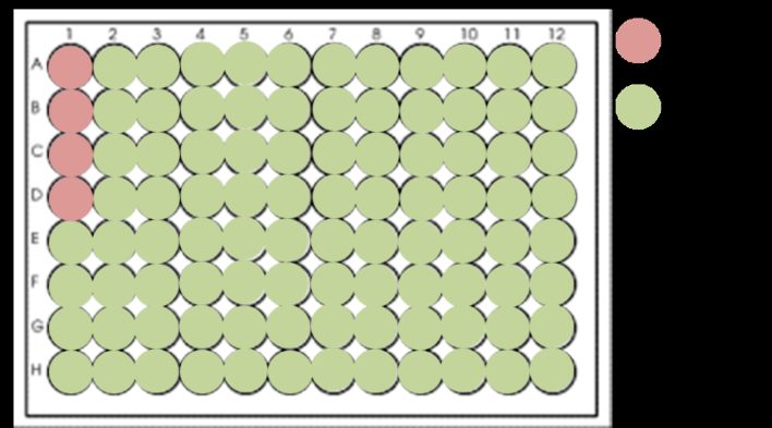
Whether or not you have counted the cells, seed the HBECs into the pre-warmed medium in the plate or flask you prepared. If you have a large quantity of cells, (i.e. a large visible cell pellet or 50,000 cells or more) split the cells evenly between 4 wells of a 12-well plate and top up the medium to 1 ml per well. For smaller harvests, limit the cells to 1-2 wells. Alternatively, you can add the entire suspension into a single well of a 6-well plate. My preference for using 12-well plates is based on the concept that HBECs prefer to maintain close-knit groups to facilitate effective cell-cell communication –dispersing them across too large an area or in too low a density reduces the chance of the culture surviving. Also, splitting the HBECs into several small wells maximizes the total number of individual cultures obtained from a single harvest.
The culture – Maintaining a healthy population
After 16-24 hours the HBECs should have adhered and started to proliferate. Any ciliated cells should disappear as they undergo a process of de-differentiation, thanks to their new environment submerged in medium. As soon as the cells have adhered, replace the medium with fresh medium frequently – every 2-3 days, or a minimum of 3 times weekly, is ideal, but the cells will only benefit if you choose to replace the medium even more frequently than this. If all goes well, the culture should grow into a pure population of de-differentiated HBECs. Under a light microscope the culture should have a tightly packed and relatively uniform “cobblestone” appearance, and should be split into more cultures when 80-100% confluent using a trypsin-based solution to gently detach the cells.
If you’re studying epithelial cell function, you simply can’t beat working with a healthy culture of primary human epithelial cells.
The next step is to get them to differentiate – but that’s a whole other ball game and deserves an article of its own.
You made it to the end—nice work! If you’re the kind of scientist who likes figuring things out without wasting half a day on trial and error, you’ll love our newsletter. Get 3 quick reads a week, packed with hard-won lab wisdom. Join FREE here.

Put this article into practice
Choose a free resource to help you move forward

POSTER
Antibiotics Reference Guide
PROTOCOL
Chemically Competent Cells Protocol