A flow cytometer is a device used to illuminate objects and capture and quantitate light emitting from these objects. The “objects” are normally single cells dispersed in a medium, but could very well be polystyrene beads, cell fragments or debris, or even large molecules.
So, What’s in the Box?
Using your highly tuned powers of deduction, you may have already figured this out from looking at the name of the technology – flow cytometry. You’ll note the roots visible in the words; namely ‘cyto’ – or cell; ‘metry’ – meaning to measure; and ‘flow’ – pointing towards the fluid-like travel through the instrument. Thinking about the technology in these terms makes the big black box less intimidating. However, in order to understand things a bit more clearly, let’s delve deeper and take a look inside the box.
The Orchestral Sections
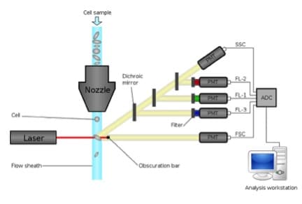
The components of a typical cytometer are separated into 4 main groups; Fluidics, Optics, Electronics, and Analysis. Each of these components works together to allow individual measurements on each cell that passes through the system. The resulting concert is a flow cytometer.
Controlling the Speed of Light
Let’s start with the Fluidics. You’ve isolated your cells and suspended them in an appropriate buffer. You’ve stained them with fluorescently coupled antibodies and/or other functional dyes, and now you’re ready to make some measurements. The purpose of the fluidics on any cytometer is to carry your cells through the focal point of the laser beam(s) at a given rate (anywhere between a couple of microliters up to 1 milliliter per minute).
The sample probe, tubing size and orifice of the sensing chamber all dictate how large a cell (or clump of cells) can pass through the instrument without clogging it (typically in the range of 50 – 250 microns). Another important job of the fluidics is to ensure the cells line up, single-file, as they pass through the focused laser beam. This is achieved in many ways, but is most commonly done using a sheath fluid (saline) that surrounds the sample stream constraining it into a narrow core.
Running at lower volume flow rates yields a narrowing of the core and more uniform illumination of the cells, whereas running at a higher volume flow rate yields a widening of the core and a variable illumination of the cells. The results of such a widening of the core stream can be seen on the software display as a spreading of the data (i.e, reduced resolution).
An ‘Optical’ Window into the Big Black Box
The Optical system illuminates the cells with one or more laser beams and provides the optical collection path to split the emitting photons into appropriate segments of the spectrum. Lasers come in a variety of wavelengths and a single cytometry system could have as many as seven different light sources.
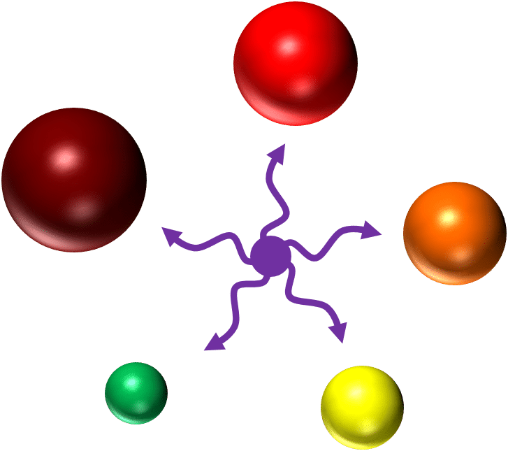
The use of filters separates the light coming from the cell and steers the photons towards a photon detector. The filters are selected and arranged according to commonly used fluorochromes. Therefore a typical system probably has filters capable of isolating green photons from fluorescein isothiocyanate (FITC) or green fluorescent protein (GFP), sending them towards the “FITC” detector, while sending orange photons from phycoerythrin (PE) to a separate detector. Of course, the ability to completely segregate FITC and PE photons from one another is not perfect. As such, flow cytometry software has a way to compensate for the inherent overlap between the emission spectra of multiple colors. But that’s a-whole-nother topic! We’ll cover spectral overlap and compensation in an upcoming article.
As the light hits the photon detector (typically a Photo Multiplier Tube (PMT)) it sends an electric current, proportional to the amount of incident light collected, to the electronics system. There is a PMT, and subsequently a current pulse, for each signal you are trying to detect.
Cell Size Matters Too!
In addition to fluorescence signals, cytometers can collect scattered laser light to glean some crude morphological information about the cells. Forward and side scattered light (FSC and SSC) can provide information as to the relative size and internal complexity of an object, respectively.
Configuration is the Key
The most important thing to be aware of on any system is which lasers are installed and what filters are upstream of each detector. This information is collectively referred to a cytometer’s optical configuration and is something that should be readily accessible to anyone operating the instrument.
The Digital Era
It is then the responsibility of the Electronics to convert this current of variable amplitude coming from each PMT, for every cell, into a number that can be utilized by software. There are various methods for performing this step, but the basic concept is the same.
The speed of the electronic components dictates how many events per second can be acquired (up to 10’s of thousands per second), whereas the bit size of the components dictate the range of signals that can be displayed simultaneously, usually spanning about 4 decades on a logarithmic scale.
The actual intensity values resulting after the electronics convert the analog signal to a digital value are always going to be relative to something. It is for this reason that negative or unstained controls are run with your stained samples in order to judge a relative increase in fluorescence signals beyond background. Each value, in every parameter, for every cell is written to a data file using the Flow Cytometry Standard, or .fcs.
It’s All in the Analysis
All this brings us to the final component of a flow cytometer; Analysis. In addition to controlling the various components of the hardware, flow cytometer software allows you to do some population analysis and gating. By drawing regions around populations with a specific phenotype (e.g. positive for FITC and negative for PE) we can get both statistical information (frequencies or relative fluorescence intensities) and look at nested subsets in a hierarchical fashion (a 3rd or 4th parameter of our FITC+, PE– population). The values for any of the parameters can be displayed on frequency histograms or correlated with other parameters in a scatter or contour plot. The only limitation here is your creativity.
So, there you have it; a crash course in how a flow cytometer works. Of course, this overview merely scratches the surface of the intricacies of a flow cytometer. However, knowing these simple basics will allow you to approach almost any instrument on the market and have some knowledge of what’s going on inside the box. Next time you find yourself face-to-face with that strange looking box with a computer tethered to it, step towards it with confidence because now you know what secrets lie beneath.
Originally published on January 21, 2013. Updated and revised on March 18, 2016.


