If you read my previous article, you may have given IHC a shot. The chances are if you’re reading this one, it may not have worked. But don’t fret, help is at hand! This article contains a few common pitfalls and problems associated with IHC, and what you can do to try and get the stain you’re looking and hoping for.
Problem: Everything just looks blue; I’m not getting any brown staining on my slide!
Possible solutions: Going through the whole staining process and getting nothing can be pretty disappointing. Luckily there are plenty of things you can try changing to fix the problem:
- Is everything fresh? Everything should be stored according to the manufacturer’s instructions. For IHC reagents, this usually means either at 4°C in a fridge, or at -20°C in a freezer. Avoid leaving things out on your bench for longer than necessary, and minimize any freeze-thaw problems by aliquoting your reagents out the first time you use them.
- Problems with your primary:
- Are you using the appropriate concentration? If your primary is too dilute you won’t get much (or any!) signal. Try a range of concentrations (maybe a 1:10, 1:100, 1:1000 dilution series) to optimize the protocol. Similarly, try incubating the antibody on the slide for longer.
- Is it cold in your lab? This might seem an odd thing to ask, but I’ve had frustrating stains in the past where one day they worked and the next they didn’t despite everything being done the same… the only variable I could account for was the sun coming through the window some days and warming up the lab. Your antibodies are as susceptible to temperature change as you are! Try adjusting your thermostat (if you have one), finding a constant temperature room, or borrowing somebody else’s lab for an hour.
- Is it suitable for IHC? Not all antibodies are created equal. Some work better on blots than stains, and some are good all-rounders. Make sure your antibody is indicated for IHC, or see if you can find some record in your literature searches for it having been used in IHC before.
- Are you using the right secondary? Double-check to make sure you’re using the right one for the species your primary antibody was raised in!
- Has your slide properly dewaxed? Some manufacturers will embed their tissues in high melting point paraffin which won’t shift if you put it straight into Histoclear. In these cases, what I find generally works is if you place the slide in an oven at 60°C for an hour, and then IMMEDIATELY (and I mean immediately, no desperate sprinting across the lab, though – have your Histoclear nearby) plunge it into the Histoclear and shake it. Then continue through the rest of the dewaxing.
- The hydrogen peroxide step to block endogenous peroxidase activity may be damaging your antigen. In this case, incubate with your primary first, followed by the hydrogen peroxide.
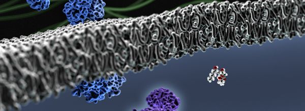
- You may be having a problem with antigen masking. Sometimes chemicals used in the fixation process (particularly formalin) can cause proteins to cross-link or lose their solubility, which make antibody binding difficult (See Fig 1). A simple method to overcome this is to (after having passed the slide through the first series of Histoclear and ethanol and washed it) submerge the slide in citrate buffer (10mM citric acid in 1 litre of distilled water, pH 6) in a coplin dish, and heat on high in a microwave for 5 minutes (it will boil… don’t worry, that’s normal!). Top up the citrate buffer and heat on high for 5 minutes again, and then leave to cool for about 20 minutes and wash twice in 1X PBS. Continue with the protocol as normal after that.
- If you try all these solutions and still get nothing, then maybe your tissue simply doesn’t express this protein much. Use some common sense here though… obviously if you’re working on some kind of prolific protein like tubulin or actin this is pretty unlikely! Have a look on the Human Protein Atlas to get an idea of what level of protein expression is found in humans.
Problem: EVERYTHING is brown!
Possible solutions: When everything goes brown, the tissue is called “toasted” (sounds yummy… but don’t eat it). It’s basically giving you too strong a background signal, so you need to try and improve your signal (brown) to noise (blue haematoxylin) ratio.
- Primary antibody problems:
- Are you using too much primary? Often I find this is the worst culprit for causing toasted slides. Try diluting it further.
- Are you incubating for too long? Or is the lab too hot? Try adjusting incubation time and the room temperature.
- Are you blocking the slide as you incubate with the primary? Blocking with 1% BSA in PBS helps to reduce non-specific background binding. If this still isn’t helping, perhaps introduce a separate blocking step where you incubate with 5% BSA for an hour before adding the primary.
- Have you carried out the hydrogen peroxide step? This is important. The hydrogen peroxide will block any peroxidase enzyme that is naturally found in the tissue which would otherwise react with the DAB chromagen at the end stages of the protocol and give a false-positive.
- Reduce incubation times for your secondary antibody and streptavidin-HRP complex. I find a lot of kits only specify 20-30 minute incubations, so anything longer than this might be causing you problems.
Problem: My tissue looks cracked/glassy/I’m having trouble focusing on it under the ‘scope
Possible solutions: This is often to do with tissue preparation and how carefully you treat it during the protocol.
- Cracked-looking tissue is often caused by it drying out during the protocol. It is VERY important that you don’t let this happen. Not only does it affect the histology of your sample, but it may also affect antibody binding. If you find yourself stuck at any point in the protocol, keep the slide immersed in 1X PBS so that it doesn’t dry out. If you’re having trouble with liquid moving around on your slide during incubations, then you can buy something called a PAP Pen. It’s basically a felt-tip marker that draws a thin hydrophobic film around your tissue and helps stop your reagent shifting around everywhere. Very handy.
- Glassy-looking tissue could be caused by the coverslip sliding around on top of the tissue after you’ve mounted it. The tissue is still delicate, so try and avoid sliding the coverslip about, and leave them to dry horizontally (flat on the table). Glassy tissue may also be caused by there being residual wax and contaminants on your slide. Check that it dewaxed properly to start with and refresh all your Histoclear and ethanol tanks.
- If you’re having trouble focusing, it may be that the tissue is cut a bit thick. If this is the case, you’ll be shifting focus through layers of cells and will struggle to find a good plane for observation. If you’re cutting the tissue yourself, then try and cut thinner sections (I generally use tissue at about 3µM in thickness), and if you’re buying, see if the company offers different thicknesses of tissue or if you can find better elsewhere. If you can’t remedy this, then it’s something you’ll have to put up with I’m afraid. The problem will be worse at higher magnification, so if you can, try and stick to 10x or 20x lenses.
Hopefully, these tips will help you out. I know it can seem like there’s a lot that can go wrong, but with a little perseverance, you’ll get a useful (and nice-looking) result! But this is only the beginning. Never underestimate the power of Google in helping you find other solutions. For example, there are a number of ways to carry out antigen retrieval, not just the one mentioned here (this is just the most commonly used). Make sure you find the protocol that’s right for you and your tissue!
Want to know more about histology? Visit the Bitesize Bio Histology Hub for tips and tricks for all your histology experiments.


