
What Is Inside a Coulter Counter
Are you an immunologist or lab personnel dealing with counting tons of cells? Then you must have come across the elusive Coulter Counter! This instrument is famous for counting thousands of cells in just a few seconds, making it possible to better plan for experiments. For this reason, you might love using it, but do you know the principle behind it? Read on to know a little history of the instrument’s technology and its working principle.
How It Was Developed
Before we dive in deep, we need to know that a hemocytometer and a Coulter counter are completely different. Also known as a counting chamber, cell counting with hemocytometer is manually done under a microscope. The Coulter counter is based on sophisticated technology using voltage and electricity to determine the cell count.
Before World War II, medical technologists spent many hours counting red blood cells using the hemocytometer (commonly known as a counting chamber), but this method resulted in poor accuracy and repeatability. After the atomic bombing of Hiroshima and Nagasaki, Dr. Wallace H. Coulter understood the need for a simple, rapid, and accurate approach to counting red blood cells of large populations, which is an index of recovery from radiation exposure. Soon, Dr. Coulter and his brother developed and perfected their Coulter method and this principle quickly changed the dynamics of cell counting. [1]
What Is the Coulter Principle?
When cells suspended in a low concentration solution pass through a small opening separated by two electrodes, they cause a change in the resistance in the electric circuit. The change in resistance is proportional to the particle volume displaced in the small opening (Fig. 1). Resistance change plotted against time gives us voltage peaks. The number of peaks is equal to the number of particles and the height of the peak indicates the size of the particle.
The majority of cell counting instruments today are based on the Coulter counter principle. These instruments take only a few seconds to analyze more than 10,000 cells, ranging between diameters of 0.4 µm and 1600 µm.
Choose a free resource to help you move forward
DOWNLOAD
Culture Contaminants Reference Card
PROTOCOL
Chemically Competent Cells Protocol
How Does the Coulter Counter Work?
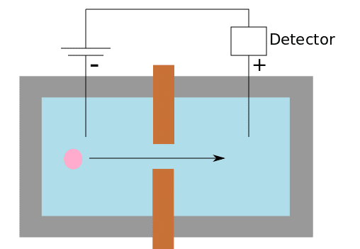
The Coulter counter instrument has the following basic construction. A beaker filled with a low concentration electrolyte with the particles of interest has a negative electrode (anode). A small tube with the positive electrode (cathode) is placed in this beaker. This tube has a small opening which acts as a “sensing zone”(Fig. 2).

This entire setup works as a closed circuit when an electric field is applied. When particles pass through the sensing zone, the volume of the electrolyte equal to the volume of the particle is displaced. This leads to a change in resistance. The resistance change plotted against time gives us voltage pulses that provide information on the number and size of the particles.
Applications of The Coulter Method
The Coulter method is widely used not only in medical and research labs, but also in many other industries because of the following advantages:
- Independent of optical and chemical properties of the particle.
- Does not require colorimetric or fluorescence.
- Can be used for any particle that can displace liquid.
- Lower power consumption.
- Detects individual particles.
Variations of the Coulter Principle
Modifications to the Coulter principle have given rise to different variations in flow cytometry and microfluidics. For example, a new method was developed to measure specific protein concentrations using the Coulter method in a microfluidic environment. Since this is a label-free technique, [2] it can be used to detect different biopolymers like DNA, protein, and blood cells. [3]
Moxi Go II is a combination of a Coulter counter and a flow cytometer, making it possible to determine the size of the cell and its primary fluorescence at the same time. This instrument also distinguishes between dead and live cells.
Many instruments have an option to change the size of the sensing zone based on the size of the particle of interest. Izon has another variation of the instrument, in which the small tube is replaced with a plastic membrane having an aperture. This membrane can be stretched or relaxed to open or close the aperture, giving a higher dynamic range of analysis.
Conclusion
Coulter counter instruments give rapid, reliable, and accurate cell counting readings. These instruments have changed the dynamics of cell counting in medical and research industries, especially in the hematology wing. The only disadvantage of the Coulter method is not being able to determine the viability of the cell, but this didn’t matter for other industries to analyze particles like pigments, clays, minerals, and other materials.
Did you ever use a Coulter instrument or its advanced variations? Share your experience in the comments below to help your fellow scientists!
References
- Graham MD. The Coulter principle: Imaginary origins. Cytometry 2013; 83(12):1057–1061. doi:10.1002/cyto.a.22398.
- Rodriguez-Trujillo R et al. Label-free protein detection using a microfluidic Coulter-counter device. Sensors and Actuators B: Chemical 2014; 190:922–927. doi:10.1016/j.snb.2013.09.038.
- Zhang H et al. Methods for counting particles in microfluidic applications. Microfluidics and Nanofluidics 2009; 7(6). doi:10.1007/s10404-009-0493-7.
You made it to the end—nice work! If you’re the kind of scientist who likes figuring things out without wasting half a day on trial and error, you’ll love our newsletter. Get 3 quick reads a week, packed with hard-won lab wisdom. Join FREE here.


