At Bitesize Bio, we share a lot of troubleshooting tips for RNA and genomic DNA extraction because almost everything we do in molecular biology requires nucleic acid isolation as the very first step. These days, most labs use commercial nucleic acid extraction kits based on spin-column technology.
Genomic DNA extraction kits are generally much easier and faster to use than traditional methods and don’t require significant expertise. The downside, however, is that troubleshooting may be difficult if you don’t understand what is in your kit’s black box!
In this article, we will go through the principles of nucleic acid extraction kits step-by-step. We will also look at some common issues with silica columns that you can overcome with a few simple tips!
A Step-by-Step Guide to Nucleic Acid Extraction Kits
Step 1: Cell Lysis
Lysis formulas may vary depending on whether you want to extract DNA or RNA. Generally speaking, lysis buffers contain a high concentration of chaotropic salts. Chaotropes have two important roles in nucleic acid extraction:
- They destabilize hydrogen bonds, van der Waals forces, and hydrophobic interactions, leading to destabilization of proteins, including nucleases;
- They disrupt the association of nucleic acids with water, thereby providing optimal conditions for their transfer to silica.
Chaotropic salts include guanidine HCL, guanidine thiocyanate, urea, and lithium perchlorate.
In addition to chaotropes, a detergent is often present in the lysis buffer to aid protein solubilization and cell lysis.
Enzymes may also feature here, depending on the sample type. The broad-spectrum serine protease Proteinase K is very efficient in digesting proteins away from nucleic acid preparations. Proteinase K works best under protein denaturing conditions (i.e., in denaturing lysis buffer).
Another popular enzyme here, lysozyme, does not work under denaturing conditions and will be most active before the addition of denaturing salts.
Bear in mind that lysis for plasmid isolation is very different from lysis for RNA or genomic DNA extraction because plasmids must be separated from genomic DNA first.
The addition of chaotropes will release all types of DNA at once, losing the ability to differentiate small circular DNA from high molecular weight chromosomes. Therefore, in plasmid preps, the chaotropes are not added until after cell lysis. For additional reading, check out these great articles on alkaline lysis and plasmid and genomic DNA extraction.
Step 2: Purification – Binding Nucleic Acids to the Column
As discussed above, chaotropic salts are critical for lysis and binding to the column. The addition of ethanol (or sometimes isopropanol) will further enhance and influence the binding of nucleic acids to the silica.
Spin columns contain a silica resin that selectively binds DNA (or RNA), depending on salt conditions and other factors influenced by the extraction method. The result is a high-quality material for cloning, long-range sequencing, and long-read sequencing, to name a few potential applications.
Note that the percentage and volume of ethanol used are important. Too much ethanol will bring down degraded material and small species that will influence absorbance at 260 nm (A260 readings). On the other hand, too little ethanol may impede the washing of the salt from the membrane.
Fortunately, the amount of ethanol added will be optimal for the nucleic acid extraction kit you are using. However, if you suspect that degraded DNA is inflating your A260 readings, you can consider re-optimizing the ethanol concentration.
Another useful tip is to save the flow-through and precipitate it to see if you can find your lost material. If you used an SDS-containing detergent for lysis, try using NaCl as a precipitant to avoid detergent contamination of your nucleic acids.
Step 3: Washing
After centrifuging your lysate through the silica membrane, the desired nucleic acids should be bound to the column, and impurities such as protein and polysaccharides should be in the flow-through. However, the membrane will contain protein and salt residues.
At this point, plant samples will likely contain polysaccharides and pigments, while for blood samples, the membrane may be slightly brown or yellow in color. The wash steps remove such impurities.
There are typically two wash steps, although this varies depending on sample type. The first wash will often include a low concentration of chaotropic salts to remove residual proteins and pigments. This is always followed by an ethanol wash to remove the salts.
If the sample didn’t contain a lot of protein starting out (e.g., plasmid preps or PCR clean-ups), an ethanol wash is sufficient.
Removal of the chaotropic salts is crucial to getting high yields and purity. Some kits actually recommend two ethanol washes. Residual salt will impede the elution of nucleic acid, resulting in poor yield, high A230 readings, and thus low A260/230 ratios.
Step 4: Dry Spin for Ethanol-free DNA and RNA
Most protocols include a centrifugation step after washing to dry the column of residual ethanol, and this step is essential for a clean eluent. Subsequent addition of 10 mM Tris buffer or water to the membrane will hydrate the nucleic acids, thus eluting them from the membrane.
Residual ethanol on the membrane at this point will prevent full hydration and elution of nucleic acids.
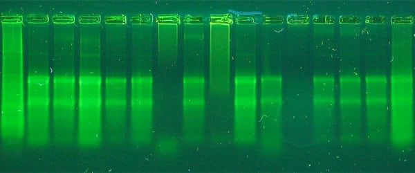
You will not be able to see ethanol on a spectrophotometer, but a good indicator of its presence is samples that will not sink into the wells of an agarose gel, even in the presence of loading dye. Another indicator of ethanol contamination is samples that don’t freeze at -20°C.
Step 5: The Final Frontier – Elution
The final step in the DNA extraction protocol is the release of pure DNA or RNA from the silica.
For DNA extraction, 10 mM Tris at pH 8-9 is typically used. DNA is more stable at a slightly basic pH and will dissolve faster in a buffer than in water. This is true even for DNA pellets.
Water tends to have a lower pH of 4-5, and high molecular weight DNA may not completely rehydrate in the short time used for elution. For maximal DNA elution, allow the buffer to stand in the membrane for a few minutes before centrifugation.
For applications requiring intact high molecular weight DNA, such as long-range sequencing and long-read sequencing, elution buffer is the best choice.
RNA, however, can tolerate a slightly acidic pH and dissolves readily in water, making this the preferred diluent.
What Can Go Wrong with Nucleic Acid Extraction Kits?
Low Yields
If you experience lower yields than you expect, there are many factors to consider. It is often a lysis issue, with incomplete lysis being a major cause of low yields. Incorrect binding conditions are another possibility. Make sure to use fresh, high-quality ethanol (100%, 200 proof) to dilute buffers and for the binding step.
Low-quality ethanol or old stocks may have taken up water, skewing the actual working concentration. Remember that if you make your wash buffer incorrectly, you may be washing away your extracted DNA or RNA!
Low Purity
If the extract is contaminated with protein, you may have started with an excess sample, increasing the risk of incomplete solubilization. If the extracts have poor A260/230 ratios, the issue is usually residual salt after binding or inadequate washing. Be sure to use the highest quality ethanol to prepare wash buffers, and if the problem continues, perform an additional wash step.
Impurities
Environmental samples are especially prone to impurities because humic substances solubilize easily during extraction. Such substances often behave similarly to DNA during the extraction process and are difficult to remove from the silica column. For samples prone to impurities, specialized techniques exist to remove interfering protein and humics prior to column binding.
Degradation
This is a greater concern for RNA than DNA extraction, and you can find specific advice on troubleshooting RNA extraction here. RNA degradation often occurs due to improper sample storage or inefficient lysis, assuming, of course, samples are eluted with RNase-free water.
For DNA extractions, degradation is not a huge issue if PCR is the desired application, but if you were hoping for intact high molecular weight DNA for long-range sequencing applications you should ensure to not be too harsh when lysing your sample!
PCR Clean-up Special Considerations
PCR cleanup isn’t a DNA extraction technique per se, but it is worth a mention here. Typically, PCR products are cleaned up by adding 3-5 volumes of salt per volume of the PCR reaction, followed by centrifugation of the mixture through a spin column.
Although a failed clean-up is often caused by an unsuccessful PCR, it is worth saving your flow-through after column binding. If a strong PCR band didn’t make it through the column, chances are it is in the flow-through. You can always rescue it and clean it up again.
FAQs
Q. What are some specific indicators or signals that might suggest incomplete lysis during the nucleic acid extraction process, and how can these be distinguished from other issues like contamination or degradation?
A. Incomplete lysis during nucleic acid extraction can manifest through various indicators such as lower-than-expected yields, incomplete solubilization of proteins, or poor A260/230 ratios.
These indicators suggest that certain components of the sample may not have been fully lysed or solubilized during the extraction process.
Distinguishing incomplete lysis from other issues like contamination or degradation requires careful analysis of the extraction protocol, including the lysis buffer composition, incubation conditions, and the presence of any interfering substances.
For instance, if the A260/230 ratios are poor, it might indicate residual salt after binding or inadequate washing, rather than incomplete lysis. To address incomplete lysis, optimizing the lysis buffer components, and incubation time, or employing additional mechanical or enzymatic lysis methods may be necessary.
Q. The article mentions specialized techniques for removing interfering substances like humic substances from environmental samples prior to column binding. Could you provide more details or examples of these specialized techniques?
A. Specialized techniques exist for removing interfering substances like humic substances from environmental samples in nucleic acid extraction kits.
These techniques are designed to specifically target and remove substances that may co-purify with nucleic acids, thereby improving the purity of the extracted material.
Examples of such techniques include using specialized extraction buffers containing chelating agents such as EDTA to selectively bind and remove cations. Additionally, pre-treatment methods such as differential centrifugation or filtration can help remove larger particulate matter from environmental samples before nucleic acid extraction, reducing the likelihood of interference during the binding step.
Q. Are there any common issues or challenges unique to specific types of samples, such as tissue samples or samples with high lipid content, that are not addressed in the general troubleshooting tips provided?
A. Specific challenges may arise with certain types of samples, such as tissue samples or those with high lipid content, which are not addressed in detail. Tissue samples, for example, may require additional mechanical disruption or enzymatic digestion steps to ensure complete lysis and release of nucleic acids.
Samples with high lipid content may pose challenges during purification due to the presence of lipids that can interfere with nucleic acid binding to the column matrix.
Addressing these challenges may involve modifying the lysis buffer composition, optimizing purification protocols, or utilizing specialized kits designed
Go Forth and Use Your Nucleic Acid Extraction Kits with Confidence
As scientists, we often want to be able to troubleshoot without asking for outside help. This article should clarify some of the science around silica spin filter technology in many nucleic acid extraction kits allowing you to troubleshoot in no time.
If all else fails, you will have done your homework by the time you call for technical support, and you should reach a resolution much faster, even if that is a free replacement DNA extraction kit!
Do you have any comments or questions about how nucleic acid extraction kits work? Leave a comment below.
Originally published June 28, 2010. Reviewed and republished May 2021 and March 2024.


