How Histology Slides Are Prepared
Ever wondered what magic happens to turn your samples into histology slides? Find out the 5 simple steps for histology slide preparation.
Join Us
Sign up for our feature-packed newsletter today to ensure you get the latest expert help and advice to level up your lab work.
Search below to delve into the Bitesize Bio archive. Here, you’ll find over two decades of the best articles, live events, podcasts, and resources, created by real experts and passionate mentors, to help you improve as a bioscientist. Whether you’re looking to learn something new or dig deep into a topic, you’ll find trustworthy, human-crafted content that’s ready to inspire and guide you.

Ever wondered what magic happens to turn your samples into histology slides? Find out the 5 simple steps for histology slide preparation.

Discover what hematoxylin and eosin staining is used for and how it works, in this concise guide.

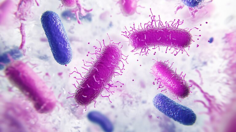
The Gram stain is another commonly used special stain in the histology lab. Why use a Gram stain? The Gram stain is a type of differential staining technique which represents an important initial step in the characterization and classification of bacteria using a light microscope. It is named after a Danish scientist, Hans Christian Gram,…

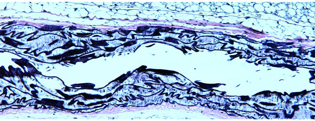
If you want to visualize elastic fibers in your sample, you need to use Verhoeff-van Gieson stain. Find out more about this stain, including how to use it.

Discover the magic of toluidine blue – a polychromatic dye that changes color depending on which tissue component it is staining.

Get introduced to some of the special stains for histology and learn some top tips for getting great results.
What Does Oil Red O Stain? Oil Red O (‘ORO’) is used to demonstrate the presence of fat or lipids in fresh, frozen tissue sections. Introduced by French in 1926, ORO is a fat-soluble diazo dye, and is classified as one of the Sudan dyes which have been in use since the late 1800s. Like…

Want to detect iron in your samples? You need Prussian blue! Discover the incredible sensitivity of this stain and how to use it.

Discover interesting facts about Congo red and it can help us understand Alzheimer’s disease.

Acid-fast stain (AF) is a special staining technique used in the histology lab. Discover which bacteria this stain detects, the history behind it, and how it works.

Gomori’s methenamine silver is a special histology stain for detecting fungi. Find out how and why you might want to use this stain in the lab.

Need to stain Gram-negative organisms? You should consider the Warthin-Starry stain.

Periodic acid-Schiff (PAS) is a commonly used special stain in the histology lab. Find out more about what this stain detects and how to use it.
Have You Tried Trichrome? The trichrome stain is one of the most commonly used special stains in every histology lab. The pedantic meaning of the word trichrome is “three-coloured”, referring to how the technique differentially stains tissue samples in three colors. However, the term is now actually used to describe any staining method using two…

It’s unclear exactly how the term ‘Special Stains’ first arose in the world of histology, but it refers to empirical and histochemical staining techniques that significantly contributed to the advancement of histology in the late 19th century. In a nutshell, these stains are ‘Special’ because they are not routine – simple as that. Therefore, Special…

I recently introduced you to the concept of polarising microscopy. Naturally, if evaluating refractile material is an everyday part of your research, it is definitely worth investing in a professional polariser modification for your microscope. But if you only use a polariser occasionally, this might not be the best use of your lab’s money. In…

Polarising microscopy involves the use of polarised light to investigate the optical properties of various specimens. Although originally used predominantly in the field of geology, it has recently become more widely used in medical and biological research fields too. Polarising light microscopy is a contrast-enhancing technique to allow you to evaluate the composition and three-dimensional…

Although the microscope is probably the most commonly used biological instrument, it is frequently used improperly. The rate-limiting step to getting high quality microscopic images is illumination of your specimen. When you examine a specimen under the microscope, the intensity and distribution of light must be clear and equal to enable you to evaluate all…

If you’re starting your PhD or post-doctoral work, chances are you’ll need to use a light microscope at some stage during your research. Some of you may be seasoned microscopists. For many of you though, this might be the first time you’ve ever plugged in a microscope, or at least the first time you’ve used…

The eBook with top tips from our Researcher community.