If you’re starting your PhD or post-doctoral work, chances are you’ll need to use a light microscope at some stage during your research. Some of you may be seasoned microscopists. For many of you though, this might be the first time you’ve ever plugged in a microscope, or at least the first time you’ve used one since some long-forgotten undergraduate histology course. Yet somehow you’re expected to just dust off that old ‘scope in the corner of the lab, and start using it as if you’re an old hand. Have no fear – help is at hand! In today’s article, I’m going to go over the basics of the light microscope.
The anatomy of the light microscope
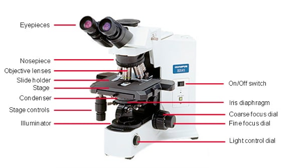
To begin with, I’d like to introduce you to the parts of the microscope – basically the bits that might be useful for you to know about. No optical physics required!
- Microscope Slide: The glass slide that contains your specimen for examination.
- Cover Slip: A smaller, thinner, flat piece of glass that lies over your specimen, and keeps it pressed flat against the slide to maintain an even thickness. This makes it easier to focus on the specimen.
- Stage: The platform where you place the microscope slide. It contains a slide holder, which is a spring-loaded clip that holds your slide firmly in place on the stage.
- Stage Controls: Dials for controlling the position of the stage. These allow you to move the slide around to bring different regions of the image into view.
- Coarse focus: This dial is used to bring the image roughly into focus.
- Fine Focus: This dial is then used to more finely tune the image into clear focus.
- Eyepieces: Also called the oculars, or ocular lenses. This is the set of lenses at the top that you look through. They collect light and focus it to produce the image that you see. They also take the image produced by the objective lens and magnify it another ten times.
- Nosepiece: A rotating carousel above the stage that contains the objective lenses.
- Objective lenses: Three or four objective lenses of different magnifications will be attached to the nosepiece – you can flip between these to view your image at different magnifications.
- Iris Diaphragm: A rotating disc under the stage that controls the intensity of light hitting the specimen.
- Condenser: Usually sits just above the iris diaphragm. Focuses light onto the specimen.
- Illuminator: The light source, which illuminates the specimen.
- Light Control Dial: Controls the brightness of light heading toward the specimen.

Viewing your slides
OK, so now that you know all the parts of the microscope, let’s look at your first slide. What do you do first?
- Switch on the microscope: I know it seems intuitive, but trust me, I’ve seen many people become frustrated at not being able to see anything, only to discover they hadn’t plugged it in or switched it to the “on” position.
- Set the slide on the stage: Allow the mechanical spring-loaded clip to grasp it in place. Make sure it’s the right way up – the cover slip should be facing upwards.
- Look through the eyepieces: Adjust the width between the eyepiece lenses to match that of your eyes (I’m keeping my fingers crossed that they’ve given you a binocular microscope, and not a monocular one!). Use the light control dial to set a comfortable light intensity. This will vary from person to person – aim for a brightly illuminated image, but not so bright that your retinas feel like they’re on fire!
- Start at the lowest magnification: Rotate the nosepiece to click the lowest magnification objective lens into place – this is the shortest lens (yours may be 2x or 4x). Use the coarse focus dial to lower the lens as close to the slide as possible, without touching it. Take care to look at the slide and lens as you do this – it’s easy to crush the slide. Once the slide and lens are close together, look through the eyepieces again to focus. Use the coarse focus dial first – turn it slowly in the opposite direction than you previously did (i.e. away from the slide) until you can see a clear image. Then use the fine focus dial to bring your image into sharp focus.
- Examine your specimen at progressively increasing magnifications: Once your image is in focus at low magnification, you can switch objective lenses and your image will remain mostly in focus – only fine focus adjustments may be required. Move up through the objective lenses in turn, to evaluate your specimen from lowest to highest magnification.
Hopefully, this overview has taken some of the mystery out of using your lab’s ancient light microscope. It can be a nightmare if you’re faced with using a microscope for the first time, but following these few, easy steps should ease the pain a little!
What are your tips for using a light microscope?


