Search below to delve into the Bitesize Bio archive. Here, you’ll find over two decades of the best articles, live events, podcasts, and resources, created by real experts and passionate mentors, to help you improve as a bioscientist. Whether you’re looking to learn something new or dig deep into a topic, you’ll find trustworthy, human-crafted content that’s ready to inspire and guide you.
Radioactivity is still the most sensitive detection mechanism for many macromolecules and enzymatic activities. In graduate school, I performed countless radioactive kinase assays, watching the radioactive gamma 32P of ATP get transferred to my autophosphorylating receptor of interest, and then separating my protein from free hot ATP on a gel. The gel is dried, covered in plastic wrap, and exposed to a storage phosphor screen for a few hours. The phosphor screen is then scanned with laser and the emitted light is converted into an image of radioactive bands on a gel.
For many years, I repeated this experiment, using the mysterious phosphor imaging screen to capture my results. But what is the phosphor screen? How does this floppy piece of plastic actually work? Here, I’ll dispel some of that mystery and outline the basic concepts of the phosphor screen.
Phosphor Screen Parts
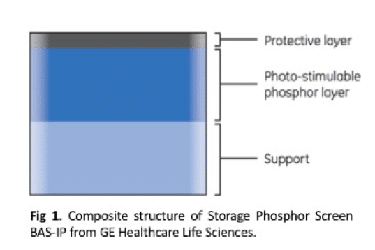
The most crucial layer of the phosphor screen is the center one (Figure 1, GE Healthcare Life Sciences). This layer contains the phosphor layer, packed with phosphor particles, which I will explain below. The important phosphor layer is shielded from physical damage with a protective layer. The base layer is for support and, depending on the manufacturer, can be a flexible plastic or a harder, more inflexible surface.
PSL: Not Just Pumpkin Spice Lattes
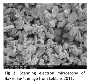
The phosphor particles are a crystal lattice of BaFBr (barium fluorobromide). This material contains photostimulable luminescence (PSL), meaning it can stably absorb energy and then release it upon laser stimulation. Photostimulable phosphors have been applied to both research and X-ray medical technology, as these materials can absorb energy from various types of radioactive emissions. Specifically, some radioactive elements emit fast gamma rays such as iodine-125, but the most popular elements in research emit less-active beta particles such as tritium (3H), carbon-14 (14C), phosphorus-32 (32P), and sulfur-35 (35S). The final crucial element involved in the chemistry of phosphor absorption is a trace amount of the divalent cation europium (Eu2+) coated onto the surface layer, which acts the “luminescent center” since it is the actual material that emits light.
Choose a free resource to help you move forward
CHEAT SHEET
SDS-PAGE Protocol Cheat Sheet
Want to avoid casting leaky and wonky gels forever? Sick of burning them by using a running buffer that’s too concentrated? Download our nifty SDS-PAGE protocol PDF that contains simple buffer recipes, gel recipes, and a neat casting protocol.
Feeling victimized by your latest Western blot? Whether you're dealing with no bands, faint bands, multiple bands, high background signal, uneven transfer, or *shudders* smearing, this handy troubleshooting card can help you gain clarity.
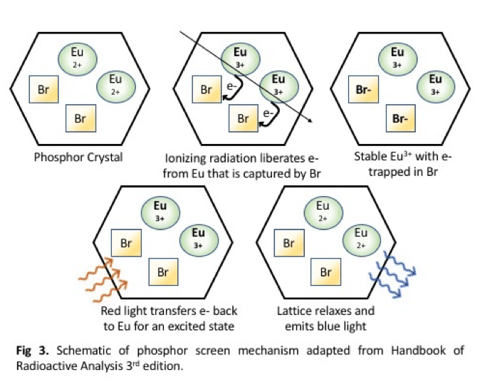
There are a few proposed models for the exact mechanism of PSL (Leblans 2011). The basic theory rests on the fact that radiation generates electron carriers that are trapped within defects in the phosphor crystal lattice. Possibly the most popular model, as it’s taught by the GE technicians of the Typhoon scanning machine, begins with ionizing radiation hitting a Eu2+ molecule. Specifically, the europium does not absorb anything, in fact the radiation actually steals an electron, making Eu3+ (Fig 2, Fig 3). The electron released interacts with bromine creating BaFBr–:Eu3+, which is a second, higher-energy stable state of the compound. These trapped electrons are sensitive to light so the exposure process takes place in a sealed cassette. To release the energy and quantify the amount of original radiation, the phosphor molecules are then hit with a red laser, to create the excited BaFBr:Eu2+* state. As the lattice relaxes to its original ground state, the europium releases a photon in the form of blue-violet 400 nm light. The light is captured by a photomultiplier tube which amplifies and converts the light into an electronic signal. The screen is then simply “blanked” by white light and can be reused thousands of times.
Mystery Dispelled
To my surprise, the mysterious phosphor screen works like an old-school television, where electron beams activate thin layers of phosphor particles within a vacuum tube. The difference lies with chemistry of the phosphor screen particles to absorb the energy in a stable manner and store it. The more you know, the more similar everything becomes.
You made it to the end—nice work! If you’re the kind of scientist who likes figuring things out without wasting half a day on trial and error, you’ll love our newsletter. Get 3 quick reads a week, packed with hard-won lab wisdom. Join FREE here.
It’s not always easy deciding whether to run electrophoresis at a constant voltage, current, or power. Here, we outline the differences to help you make an informed decision.
Wouldn’t it be great to put your nucleotide sequence into a program and get back a 3D-structure of your protein and a full description of its functions? In theory, because the protein 3D-structure is determined by the aminoacid sequence, given the right algorithm and a powerful enough computer, this should be simple. In practice, because…
Low yield in cell-free protein synthesis is usually fixable… if you know which part of the system is failing. The most common culprits are easy to address: template purity, transcription or translation inhibitors, and potassium and magnesium concentration. Check those first. But when the basics are in order and yield is still poor, the problem…
For Western blot data to be reliable, it is important that you load known amounts of sample into each lane of the gel. This is of particular importance if you are doing a quantitative blot, where you really need to be able to compare band intensity in each sample. In this article, we’ll talk about…
You use your antibody frequently, maybe even every day. You rely on it for western blotting, immunohistochemistry, FACS, ELISA, and immunoprecipitation. You’d be lost without it. But how well do you really know your antibody? Are you sure that it detects what you believe it to detect? If you have the slightest doubt, or if you have…
Purifying a new protein is no easy feat. Finding combinations of protein purification buffer, salt, detergent, and stabilizing agent to get high yields of squeaky-clean protein can become tedious. Few things are as bothersome during this process as Heat Shock Protein (HSP) contamination. But worry not, we’ve got some handy tips to avoid HSP contamination…
1-2-3 Newsletter
Get help with everything* lab-related.
*Well, everything except the washing up. That’s still on you.
10 Things Every Molecular Biologist Should Know
The eBook with top tips from our Researcher community.