Studying immune cell activation allows scientists to understand the way the body mounts a response to a specific infection, autoimmune diseases, or cancer. This knowledge plays a direct role in developing more efficacious vaccines and therapies.
When tasked with capturing information on immune cell activation, flow cytometry remains the gold standard due to its versatility, user-friendliness, and ease of data production. Dialing back the time to the 1970s, immunologist Len Herzenberg was using microscopes to study his fluorescently labelled monoclonal antibodies. 1 Unfortunately, this was time consuming and he had poor eye sight. Herzenberg realized that he could add a light source and a photomultiplier tube to Mack Fulwyler’s Los Alamos sorter and the Fluorescence Activated Cell Sorter (FACS) machine was born.
Now, the term “flow cytometry” is generally used to describe both analyzers and sorters as the term ‘FACS’ is actually commercialized.2 Flow cytometers analyze individual events and record multiple parameters at rates of several thousand events per second. The interrogation of such data sets allows for the quantification of thousands of cells in a very short period of time, resulting in statistically robust data. There are now machines available that can be used to see up to 27 different fluorochromes, which has opened up the field of immunology.
Time to go with the flow!
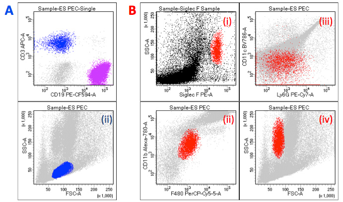
Before you begin your immune cell activation experiment, there are several parameters to take into consideration. First, you need to be able to identify the cells of interest for your assays, whether that means isolating them from a mixed cell population or using cell surface markers to distinguish them from other cell types.
Also, the method of stimulation you choose (e.g., target cells, chemical, antigen peptides) will impact how you measure the activation that occurs. For instance, a degranulation assay would be more appropriate if stimulating cells of interest with target cells (looking for cytotoxic capabilities), whereas chemical and antigenic stimulation would be preferential for proliferation studies.
Finally, be aware that activating immune cells can result in a change in phenotype, gene regulation, and possibly function.
Here are three of the most used ways to examine immune cell activation via flow cytometry.
1. Surface antibody staining
Before encountering a pathogen, immune cells exist in a quiescent/resting state. Stimulation with a foreign pathogen or exposure to a pro-inflammatory milieu leads to an intracellular signaling cascade that culminates with genetic changes resulting in alterations to cell surface proteins.3
For instance, when presented with the appropriate antigen, a naïve T cell will be stimulated to present maturation markers (e.g., CD69 and CD25) that aid in proliferation of this newly differentiated effector cell. Changes in the expression of surface proteins can be measured using flow cytometry. For even more information on cell functionality (e.g. proliferation, pro-/anti-inflammation cytokine production, cytotoxic protein production), you can also combine surface antibody staining with intracellular cytokine staining.
2. Intracellular Cytokine Staining
There are 4 main characteristics of any immune response: (1) calor (heat); (2) dalor (pain); (3) tumor (swelling); and (4) rubor (redness).4 These physical attributes are a direct result of an influx of immune cells trafficking to the infection site because of the release of cytokines from the sentinel cells that have encountered a foreign body. Cytokines (cyto = cell; kine = kinesis/movement) are small proteins that are important in cell-to-cell communication, immune cell trafficking, and setting the environment milieu (i.e., pro-inflammatory v. anti-inflammatory).
Cytokine measurement allows researchers to investigate the response to specific antigen stimulation, and the use of flow cytometry provides the ability to determine what cytokines are being produced by what specific cell type. To accomplish this, stimulated cells are first treated with an agent that blocks protein transport, such as brefeldin A or monensin5, and then fixed and permeabilized to allow for your cytokine-specific antibody to reach its target. Be sure to use an antibody directly conjugated to a “bright” fluorophore for labelling intracellular cytokines (e.g., GFP/FITC, PE, etc.) – they are often less abundant than cell surface proteins, so this will give you the best chance of detecting their presence!
3. Cell Proliferation
Following a response to a pathogen, immune cells will become activated and proliferate to increase their population numbers. This is an important point to study, particularly when trying to uncover what immune cell populations expand the most during a specific infection, or how age plays a role in cellular activation responses.
Ki-67: The nuclear protein Ki-67 is a marker commonly used to measure cell proliferation as it is upregulated during active phases of cell cycle.6 While it is incredibly easy to use in comparison to other methods, researchers often complain of high background – an important point to keep in mind during experimental set-up on the cytometer as well as during data analysis. Also, similar to intracellular cytokine staining, be sure to use an anti-Ki-67 antibody with a directly conjugated “bright” fluorophore – let’s make sure you detect proliferation!
BrdU: One method of studying proliferation is to tag the DNA that is being replicated. This was traditionally done using radio-labelled thymidine7, which can be incorporated into the DNA during replication, and then detected using liquid scintillation. However, radioactive- based assays have huge drawbacks. To get around this hazard, the thymidine analog 5’-bromo-2’deoxyuridine (BrdU) was developed.7 BrdU can also incorporate into newly synthesised DNA of replicating cells during S phase instead of thymidine. Anti-BrdU antibodies conjugated to a fluorescent tag are used to detect BrdU via flow cytometry. However, because it replaces thymidine, BrdU can cause DNA mutations.
Dye-dilution methods: Dye-dilution methods utilize the fluorescent labelling of cellular proteins with dyes such as Carboxyfluorescein N-hydroxysuccinimidyl ester (CFSE). CFSE is a cell-permeant dye that covalently couples to intracellular molecules to retain CFSE within the cell for long periods of time.8 As the cells divide so does the CFSE, resulting in each cell division to be observed as a halving of the CFSE fluorescence from the previous stage.
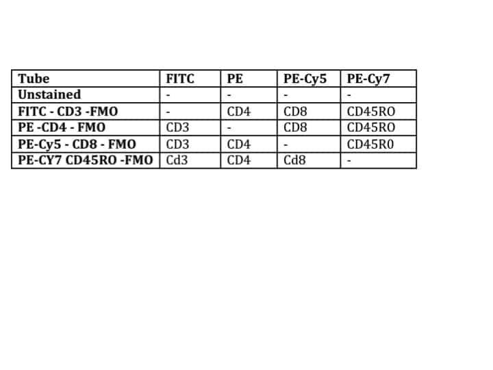
This simple assay is easy to set up and can be combined with other cell surface staining on a flow cytometer. CFSE can be very bright and therefore care needs to be used when combining with other fluorochromes to ensure that the correct controls for compensation and gating are used. Because CFSE and GFP have the same excitation/emission spectra, it is important that they are not used together in the same experiment. There are other dyes that are similar to CFSE for studying dye-dilution methods but have different excitation and emission profiles and therefore can be used in combination with GFP or other fluorescent proteins.
Flow cytometry is an important tool for studying immune cell activation under a number of conditions. Because flow cytometry permits the user to examine thousands of cells per second, it is possible to gather large amounts of data very quickly. With the advent of more affordable flow cytometry machines and more fluorochromes being available, it has never been easier to measure immune cell activation.
We founded Astarte Biologics in 2004 with one focus: to provide the highest quality immune cell products. From donor testing and consent to detailed Certificates of Analysis and fast, reliable shipping, we do everything in our power to ensure you receive viable, functional cells every time.
References
- Dangl, J.L. and Lanier, L.L. Founding father of FACS: Professor Leonard A. Herzenberg. PNAS. 2013; 110(52): 20848-20849. doi: 1073/pnas.1321731111.
- Lanier, L.L. Just the FACS. J Immunol. 2014; 193(5): 2043-2044. doi: 4049/jimmunol.1401725
- Pearce, E.L. and Pearce, E.J. Metabolic Pathways in Immune Cell Activation and Quiescence. Immunity. 2014; 38(4): 633-643. doi: 1016/j.immuni.2013.04.005
- Xiao, T.S. Innate immunity and inflammation. Cell Mol Immunol. 2017; 14(1): 1-3. doi: 1038/cmi.2016.45
- O’Neill-Anderson, N.J. and Lawrence, D.A. Differential Modulation of Surface and Intracellular Protein Expression by T Cells after Stimulation in the Presence of Brefeldin A. Clin. Diagn. Lab. Immunol. 2002; 9(2): 243-250. doi: 1128/CDLI.9.2.243-250.2001
- Scholzen, T. and Gerdes, J. The Ki-67 protein: From the known and the unknown. J Cell Physiol. 2000; 182(3): 311-322. doi: 1002/(SICI)1097-4652(200003)182:3<311::AID-JCP1>3.0CO;2-9
- Madhavan, H.N. Simple Laboratory methods to measure cell proliferation using DNA synthesis property. J Stem Cells Regen Med. 2007; 3(1): 12-14. PMID: 24693014
- Quah, B.J. and Parish, C.R. The Use of Carboxy fluorescein Diacetate Succinimidyl Ester (CFSE) to Monitor Lymphocyte Proliferation. J Vis Exp. 2010; (44): 2259. doi: 3791/2259


