In my previous article I covered different immunohistochemical staining techniques at a superficial level. In the following articles I will start to explain these technologies in a bit more detail and in which situations they should be applied.
All of the following will involve additional stages when applying them, for example- serum blocking, protein blocking, avidin biotin blocking, peroxidase blocking and so forth, but for this article (and Part 2) we will concentrate solely on the principle of the detection.
The Direct Method
The direct method utilises directly labelled primary antibodies. They may either be purchased already labelled, or can be labelled by the researcher with the label of choice using the appropriate chemistry such as HRP labelling https://www.scribd.com/doc/17410323/How-to-HRP-Label-and-Purify-Your-Antibody-No-HPLC-Needed.
It doesn’t have all the answers though
The direct method lacks some versatility as it is not as sensitive as so-called ‘sandwich’ methods.
Put this article into practice
Choose a free resource to help you move forward

EBOOK
Guide to Special Stains for Histology

POSTER
Immunofluorescence Troubleshooting Guide
Furthermore your primary antibody is now tagged with a label which may not be suitable for future applications. One final disadvantage is the limited range of primary antibodies which are directly labelled, and if you need to label the antibody in the lab then this involves a time commitment.
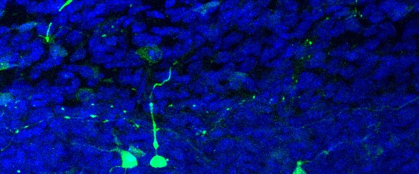
The direct method can be used for applications such as fluorescently labelling primary antibodies in co-localisation experiments where the target protein is abundant and the primary antibody has high specificity.
A current application…watch this space!
Here’s an example of a current problem where the direct method may offer a solution. A specific application in which I am considering this approach is mouse on mouse detections (‘MOM’ i.e. a mouse monoclonal antibody applied to mouse cells or tissue). Normally I would use a proprietary MOM kit, but I currently have a problem antibody! The primary antibody is giving the required staining, but is dogged by high background. Having exhausted other options, the background staining may be eliminated by labelling the primary antibody…..watch this space. In theory, this should improve the background as there will be no ‘anti-mouse’ reagents involved in the detection, but the question is, will it have the required sensitivity?
The Indirect Method
The indirect method utilises labelled secondary antibodies and has much greater flexibility than the direct method. There is a large range of fluorophore, hapten or enzyme labelled secondary reagents available to the researcher. This means that you could detect your primary antibody in a variety of ways depending on the end point that is required.
A Golden Rule – Remember It Well
The indirect method is still used extensively in flow cytometry and for western blotting as well as immunohistochemical detections. It is here that one of the golden rules of immunohistochemistry emerges; remember it well and you will never regret it. The rule is to block using normal serum from the same species as the secondary antibody.
For example, if you were using a rabbit primary antibody and a goat anti rabbit HRP secondary, you would incorporate a serum blocking step using normal goat serum before application of the primary antibody. The concentration of the blocking serum varies (between 2.5% and 20%) but the idea is to block any sites on your cell or tissue which, in this case, goat IgG would bind to. By blocking these sites with normal IgG, your labelled secondary will be unable to bind to them and will therefore prevent false positive staining.
Not entirely sensitive
If the direct method had the sensitivity of methods such as streptavidin or polymer detections it would be used much more extensively, but it is relatively insensitive in comparison to these techniques.
In part two, we’ll look at PAP, APPAP and sandwiches. Yes, sandwiches!
You made it to the end—nice work! If you’re the kind of scientist who likes figuring things out without wasting half a day on trial and error, you’ll love our newsletter. Get 3 quick reads a week, packed with hard-won lab wisdom. Join FREE here.

Put this article into practice
Choose a free resource to help you move forward
EBOOK
Guide to Special Stains for Histology

POSTER
Fluorescent Proteins Guide