Phosphorylation is one of the major post-translational modifications that regulate the activity of a protein. Around a third of human proteins are believed to be phosphorylated, and so the kinases and phosphatases that mediate protein phosphorylation are of major interest to biomedical researchers.
However detecting protein phosphorylation can be difficult, particularly from cell extracts. Phospho-specific antibodies are useful if you already know you protein is phosphorylated, and on which residue, but if there isn’t one already available making one is expensive, time consuming, and with no guarantee of success.
If you don’t have ready access to a mass spec facility, even finding if your favourite protein is phosphorylated in vivo is far from simple. Phosphorylation often causes proteins to migrate more slowly through acrylamide gels. Whilst this can allow you to monitor protein phosphorylation, it gives no indication how many times your protein is phosphorylated, and many phospho-proteins can’t be detected in this way.
Using phos-tag acrylamide to detect protein phosphorylation
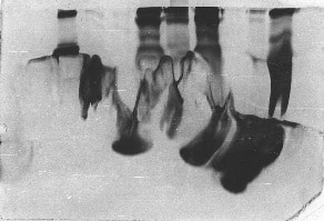
Over the last couple of years, I’ve been using Phos-tag acrylamide to detect protein phosphorylation from cell extracts. Phos-tag is a reagent developed at the NARD institute in Japan, which specifically binds phosphate groups. Phos-Tag acrylamide can be incorporated into your SDS-PAGE gels. The Phos-tag will then bind to phosphorylated proteins, selectively retarding their progress through the gel. The more a protein is phosphorylated, the slower it will migrate. Hence Phos-tag gels should, in principle, resolve different phospho-species as individual bands.
Using Phos-tag acrylamide certainly isn’t without its difficulties. For example, Phos-tag gels take much longer to set, and are more fragile than conventional SDS-PAGE gels. This is a particular concern for those of us familiar with the “acrylamide jigsaw” that inevitably results from an especially sausage-fingered attempt to transfer a gel into the blotting apparatus. If your protein of interest is very large, this becomes more of an issue, due to the low acrylamide content of the gels you have to make to resolve phospho-proteins from their unphosphorylated siblings. There are reports of high molecular weight phospho-proteins being detected on ultra-low acrylamide gels augmented with agarose, but I have never tried this.
It can also take a while to find the optimum concentration of Phos-tag acrylamide to add to your gel, particularly if you’re probing whole cell extracts. There doesn’t appear to be a definite formula for how much to add, so a certain amount of trial and error is involved. In addition, some proteins run with altered mobility on Phos-tag gels. At higher Phos-tag concentrations this includes some molecular weight markers; making detecting your protein of interest difficult if the antibody you’re using isn’t super-clean.
However, after a little optimisation, I’ve been able to generate reproducible, verifiable band-shifts to demonstrate that a protein is phosphorylated. Sadly, this reagent doesn’t tell you where your protein is phosphorylated, or the kinase/phosphatase responsible, or what the phosphorylation actually does. That’s the where the real fun starts.


