
Drosophila melanogaster, otherwise known as the common fruit fly, is one of the oldest and most powerful model systems used in biology.
Fruit flies are cheap to maintain, and have a shorter life cycle and higher fecundity than mammalian models. They also have extraordinary genetic tools with which to investigate many molecular and cellular questions. The diversity and strength of these genetic tools comes from the complex simplicity of Drosophila genetics.
Gender Determination in Adult Fruit Flies
By Chromosomes
Unlike humans, fruit flies have only four chromosomes: three autosomes, denoted 2, 3, and 4, and two possible sex chromosomes, denoted X or Y. Unlike mammals, however, it is not the absence or presence of the Y chromosome that determines gender. Rather, it is the ratio of female determinants on the X chromosome and male determinants on the autosomes.
If there is only one copy of the X chromosome in the fly, the fly is male. If there are two copies, the fly is female. And while the Y chromosome contains some genes important for spermatogenesis and sperm motility in adult male flies, it does not intrinsically determine gender. The X chromosome, however, encodes genes important to cellular function, and therefore, as you make manipulations on the X chromosome, it becomes important to determine whether you should use males or females.
Choose a free resource to help you move forward
DOWNLOAD
Hemocytometer Cell Counting Helper Pack
POSTER
Antibiotics Reference Guide
By Phenotype
Superficially, you can tell a male fly from a female fly by comparing the shape and color of their genitalia, as well as the absence or presence of sex combs on their legs. Adult male flies have dark, rounded, complex structures on the lower tip of their abdomen which comprise the genitalia (penis, lateral plate, genital arch). On the other hand, female flies have light, pointed genitalia comprised of very few visible structures. In addition to differences in the color and shape of genitalia, male flies also display a unique set of sex combs on the fourth segment of their front legs. These cells look like a row of thick bristles that appear as a dark mass on their legs and are used by male flies during copulation to grasp the female fly.
Gender Determination in Drosophila Larvae
While the superficial distinctions between male and female adult flies can be easily seen under a microscope, the difference between male and female larvae is a bit more nuanced. Fruit fly larvae are little tubes of muscle, intestine, fat, heart, brain, and trachea all encased and protected by a sturdy, yet pliable, translucent cuticle.
Some fruit fly biologists claim that female larvae are bigger than male larvae, but this distinction is not easily quantified and should not be relied on in the lab. Rather, the best way to determine larval sex is to take advantage of the translucence of the cuticle and look for the absence or presence of gonads. In male larvae, gonads are big, circular, translucent disc organs that can be seen through the cuticle of the lower abdominal segments. In female flies, these translucent discs are not present.
Example Pictures
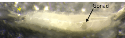
Here is an example of a pinned third instar larva. The head of the larva is denoted with a yellow star and an arrow points to the presence of a gonad. Indeed, it is a translucent, circular disc and we definitely have a male larva on our hands.
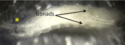
Here’s another view of a pinned larva. This time we see both the right and left gonad against the white trachea, proving again that it is a male. When the larva crawls, you’ll see these discs move along the body wall with every contraction but remain in their relative position.
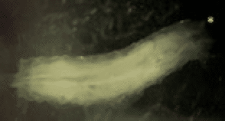
Female larvae will not show the presence of a pronounced disc. Instead, you see predominantly fat bodies and intestines.
Sexing larvae, as it is called in Drosophila larvae, is a highly nuanced skill that takes both time and practice. To double check whether or not you are doing it correctly, you can test your prowess with a mini experiment. Take some larvae, determine their genders, then place each one in their own food vial and allow to eclose into adulthood. Determine the gender of the adult and compare it with your larval determination. Hopefully they match!
Working with larvae for assays? See how to feed fruit fly larvae small molecules.
You made it to the end—nice work! If you’re the kind of scientist who likes figuring things out without wasting half a day on trial and error, you’ll love our newsletter. Get 3 quick reads a week, packed with hard-won lab wisdom. Join FREE here.
 4")


