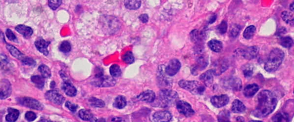
Automated image analysis uses finely tuned software to extract data from digital images. Algorithms recognize specific shapes and patterns in the images and gather quantitative information that is then used for further data analysis.
The pharmaceutical and biological research industries have benefitted greatly from this technology, which allows researchers to analyze hundreds—if not thousands—of samples in a fraction of the time that it would take a person to do the same.
How Does Automated Image Analysis Work?
Automated image analysis is one component of a larger automated system known as High Content Screening (HCS) or High Throughput Microscopy (HTM). These systems can take thousands of biological samples through the entire sample preparation, treatment, and imaging stages of an experiment. Sample imaging is performed with a digital microscope, which captures digital images of the samples. The digital images are then collected and stored before being passed on to the image analysis system.
Digital images consist of thousands of tiny squares called pixels. Each pixel has particular values assigned to it. IA uses this pixel information to determine the shape, size, and pattern of the objects that make up the image. An example of the parameters that can be quantified with automated image analysis are:
- Condition of the nucleus, cytoplasm, membrane
- Distribution and quantity of proteins
- Gene expression
The algorithms then make some advanced calculations with this shape and pattern information, eventually leading to results.
Why Use Automated Image Analysis?
Researchers dedicate huge amounts of time to data gathering. Traditionally, this would involve a person going through each sample individually, assessing and measuring the parameters of the sample, and then recording the quantitative or qualitative values. These values would then be entered into a database for data analysis. While this approach generates a great deal of knowledge and experience for the researcher, it also poses a number of challenges or difficulties, such as:
- For trials to be valid, a huge number of samples are required
- One must reproduce the results of those samples for statistical significance
- Trials, repeats, and spot-checking are time consuming for the researcher
- Human error. People are prone to making mistakes and after several hours of looking at samples, researchers may get “sample blindness.” This is where one becomes immune to what they see and the samples all start to blur into one
- When combined with tiredness or distractions, human error will also to mistakes and misdiagnosis
- People score their samples based off of their own understanding and opinion. This can lead to variation in results if more than one person is reviewing samples
With automated image analysis, you eliminate the risk of a person making a mistake or interpreting a result incorrectly. Moreover, you reduce the need for manpower.
What Are the Requirements for Automated Image Analysis?
Equipment
Image analysis requires a very powerful computer that can handle large image files. Algorithms can be complex and generate high volumes of data, so a computer that can chew through data at high speed is essential. The images and data also need to be stored, so a large memory bank is required. This is often provided as a server or other external storage device that is dedicated to imaging.
If the image analysis system is part of an automation pipeline, then the computer will need access to the storage device where the images to be analyzed are waiting. Of course, the image analysis system can also be separate from the pipeline. In this case, the user will provide the image analysis software with digital images on a storage device.
Image Requirements
Good images are key to obtaining a good data set. The samples must be well prepared—tissue should be lying flat and cells should not be too clustered. The staining should be moderate, there should be no background staining, artefacts, or aberrations. The image should be in focus, with appropriate lighting (not under- or over-exposed). Images where cells are very clustered or out of focus are not impossible to work with, although these will require intervention from the researcher.
Training
Initial training on the analysis software is most often a part of the purchased software package, and this training will prove invaluable to the users when setting up new trials or when doing spot-checking and trouble-shooting on the system.
Advantages of Automated Image Analysis
- It can extrapolate large volumes of quantitative data in a fraction of the time that manual image analysis would require.
- Minimizes human error or differences in opinion that may arise during the data collection process. People can make mistakes when tired or distracted. Software never gets tired, and gives consistent performance over time.
- While the computer performs the data collection, lab personell are free to do other work.
- An established system reduces costs, as multi-tasking is now possible, reducing personell requirements in the lab.
- The system can run overnight or even over the weekend, further saving time and human effort, all while speeding up the data analysis process.
- Once the setup is optimized, the results are reliable, and algorithms can be used indefinitely.
Disadvantages of Automated Image Analysis
- Image analysis software and algorithms can be expensive to buy. Algorithms are also brand specific, so they must be purchased from the original supplier.
- Optimization time is required to fine tune the algorithms for image analysis. Algorithms are designed with a certain task in mind, for example, to pick out individual nuclei.
- The software requires training for optimal use and accurate results. Although training is often included with the purchase of the software, any new staff will also need to be trained, either in-house or another training session needs to be purchased.
Automated image analysis is an amazing technology that is growing more and more sophisticated by the day. While the initial financial and training output can be costly and time consuming, the technology more than makes up for that with its efficiency and reliability.


