Why do I see a cloud?
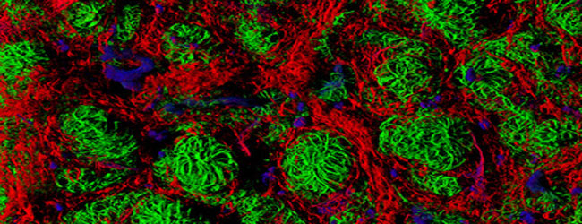
When you are looking at protein localization within a cell, have you ever wondered why you see a cloud of fluorescence rather that several individual fluorescent points? Well, light microscopy has a theoretical resolution limit of 200 nm. This means that in theory, to resolve two points as being distinct they must be at least 200 nm apart.
It’s all just a blur…
However, in reality even the best microscopes rarely approach this resolution (this includes confocal and two-photon microscopy). The problem is particularly pronounced with cellular fluorescence imaging where you have a lot of individual fluorescent points crowded together. As a result, rather than many distinct points you get an indistinct cloud of signal.
Enter Super-Resolution!
The cloud of signal may be good enough if just want to know which organelle your protein of interest localizes to, but what if you aspire for more? Where does the protein go within that organelle? Is it distributed uniformly or in distinct patches? What if you would like to see trafficking of individual proteins? Enter super-resolution light microscopy.
Too many, too close
The source of the resolution problem is that you have too many fluorescent points which are just too close together. Super-resolution microscopy gets around this by limiting the number of fluorophores that are fluorescing at a given time. As a result, the fluorescent points are much more than 200 nm apart and you can resolve the location of individual proteins.
Crisp and distinct
But wait, don’t you lose a lot of information if only a small subset of proteins are fluorescent? When you take an image with a super-resolution technique, the sample is actually scanned a number of times and information from all the scans is combined into a final image. Now rather than a diffuse cloud of signal, the final image contains crisp and distinct fluorescent points.
The alphabet soup
There are a few different super-resolution techniques that you hear a lot about: stochastic optical reconstruction microscopy (‘STORM’), photoactivated localization microscopy (‘PALM’), and fluorescence photoactivation localization microscopy (‘fPALM’). The difference between the approaches has to do with what fluorophores and microscopes are used. Let’s start with STORM.
STORM
In STORM, two lasers and a photo-switchable dye (originally Cy3 and Cy5) are used on a TIRF (total internal reflection fluorescence) microscope (Rust et al., 2006). The dye is attached to the protein of interest through antibodies and standard immunohistochemistry.
The imaging procedure goes like this: a red laser turns all fluorophores ‘off’, a green laser turns a small and random number of fluorophores ‘on’, a picture is taken, the red laser turns all fluorophores ‘off’, repeat. After a set number of repetitions (around 60) the individual images are processed and merged into a final image. This procedure works because of the photoreactive properties of the fluorophore: red light efficiently deactivates it and green light inefficiently activates it.
PALM and fPALM
The PALM and fPALM techniques use a similar imaging procedure, but instead of photo-switchable dyes they use photo-switchable protein fluorophores (such as photoactivatable green fluorescent protein (PA-GFP)) to genetically tag the protein of interest (Betzig et al., 2006; Hess et al., 2006). The difference between PALM and fPALM is that PALM uses a TIRF microscope while fPALM uses a more traditional confocal microscope.
A whole new world has opened up
All three of these techniques were originally published in 2006. There are now several more fluorophores available, as well as faster cameras that allow you to combine super-resolution imaging with live cell imaging to follow protein trafficking through the cell. Whether STORM, PALM or fPALM is right for your system will depend on the microscopes available, if you are working in a genetically tractable system and if you have a good primary antibody against your protein of interest. With these techniques the world of nanometer resolution imaging has finally opened up to light microscopists.


