Confocal Images: Expert Tips for Your Hour of Need!
Getting publication perfect confocal images can be tricky. If you are struggling or just want to ensure you’re capturing the best images possible, check out our top 7 tips for confocal imaging.
Join Us
Sign up for our feature-packed newsletter today to ensure you get the latest expert help and advice to level up your lab work.

Getting publication perfect confocal images can be tricky. If you are struggling or just want to ensure you’re capturing the best images possible, check out our top 7 tips for confocal imaging.

Continuing from our first article on lasers for confocal microscopy, we will now discuss two specialized types of lasers: lasers for two-photon excitation and tunable, white light lasers. We will also discuss the applications of the two lasers. Lasers for Two-Photon Excitation The two-photon absorption phenomenon was first described for microscopy in 1931. Here, the…

Purchasing a microscope camera is one of the most daunting tasks you might have to undertake. Before you set out to buy that camera, carefully consider your applications. Things like sample brightness or the speed of the phenomenon you are trying to capture can dictate your choices. Also, this is the time to make peace…

While using human clinical samples in your research can provide robust and heterogeneous results applicable to larger portions of the population, working with these samples presents its own set of challenges. Here are some tricks I have learned to help isolate and grow your cells of interest while eliminating stromal, blood, or other undesired contaminants….

Are you preparing to set up live cell imaging experiments? You’ve got all your cell lines, antibodies, reagents, and protocol ready. You just want to wake up in the morning and enter into that dark room. Well, think again!! As we (I mean the cell biologists) always say, happy cells mean happy life. You have…

Lasers were once called “a solution looking for a problem.” The word—which is an acronym for Light Amplification by Stimulated Emission of Radiation—used to conjure up images of deadly weapons from Sci-Fi movies and TV series. However, their increasing use in everyday life, first in CD players and then in barcode scanners and pointers, have…

Fortunately for microscopy users, measuring intracellular fluorescence has been made relatively simple through an ImageJ plugin called the Cell Magic Wand. For those of you unfamiliar with ImageJ, it’s a popular image processing program that runs on Mac, Windows, and Linux. How to use ImageJ for measuring intracellular fluorescence First of all, to begin measuring…

First of all let me say the technique of labeling tissues (immunohistochemistry, IHC), and cells (immunocytochemistry, ICC) is indeed immunoscience NOT alchemy, though at times it may certainly seem like alchemy! But to scientists inexperienced in this technique, who typically see the results of IHC/ICC experiments in the form of pretty pictures, it can certainly…
The concepts of brightness and contrast are so general, and the issues related to them so many, that it may seem strange to have a single brief article with such a title. Indeed, when we speak about brightness, we can think about the brightness of the light source, the aperture and magnification of the lens,…
When we are doing a double labelling, it makes good sense to choose two fluorophores with spectra widely spaced apart to avoid mixing. Why combine GFP with Rhodamine, when you can combine it with Texas Red?

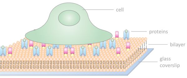
We have to rely on artificial systems to test our hypotheses and often have to come up with original set-ups to investigate specific problems. One of these creative inventions is the use of supported lipid bilayers.

What do you use if antibodies are too large for super resolution microscopy? Aptamers. These are small affinity reagents (~ 2 nm!) that interact with their target in the same way as an antibody, but without the hefty backbone attached.

When you first start out using a microscope, you might only adjust the eye pieces, objectives, and the focus controls. However, you shouldn’t overlook the microscope condensers as they are an important part of the whole optical system of a microscope.

Supported lipid bilayers are a very useful tool in many fields of cell and membrane biology. But how easy is it to make them? Bilayers can be made quite reproducibly, once you have found a reliable protocol! However, it can take some time to optimize your technique, so to increase your chances, make sure you…

Taking publication quality immunofluorescent images of can be a very time intensive, and frustrating process with hours spent capturing, processing, and putting the images into final figure format. And, if you aren’t careful, you can do a lot of work only to realize later that you need to re-image something for one reason or another….

If you want to see in real time what is going on inside your cell then you should be performing live-cell imaging. Live-cell imaging techniques allow real-time examination of almost every aspect of cellular function under normal and experimental conditions. With all live-cell imaging experiments, the main challenges are to keep your cells alive and healthy…

Do you suspect that your favourite protein is doing something really cool? But you cannot see it because your confocal microscope’s resolution is limited. Then Stimulated Emission Depletion (STED) microscopy is what you need! With the power to smash through the diffraction limit of confocal microscopy, STED opens up a whole new world of improved…

The eBook with top tips from our Researcher community.