Jennifer Redig
I have a Masters of Clinical Research, and a PhD in Molecular & Medical Genetics. However I love keeping up with a wide variety of scientific topics – making my work as a Managing Editor at BitesizeBio very enjoyable.
I am passionate about academic reform and being a mom. Connect with me on Twitter and Facebook!

Articles by Jennifer Redig
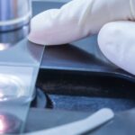
Adherent cell fixation is a crucial step in preparing cells for microscopy and imaging, ensuring that cellular structures are preserved for detailed analysis. Read our 8-step guide on how to effectively fix adherent cells to your microscope slides, including tips on sterilization, coating, and fixation methods, right here.
Discover seven common histology mistakes and how you can avoid making them when performing your experiments.
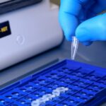
If you need to copy, sequence, or quantify DNA, you need to know about PCR. Read our guide to the PCR process, and discover tips to help you avoid the most common PCR pitfalls.

Are you having problems with tissue sectioning? Follow these 10 tissue sectioning tips to create the perfect tissue section every time without stressing out.
Would you eat your spaghetti dinner without a plate? No, of course not! It would make a big mess and be ugly to look at. Instead you NEED something to put that spaghetti on, to contain it, to keep it clean, to make it look nice – a bowl, a plate, SOMETHING! By the same…

The biggest source of PCR contamination is aerosolized PCR products. How do you fix PCR contamination and avoid it in the future?
Yup, we really are in the digital age…even the pathology is digital. Facebook, Instagram, Tumblr. It has never been easier to take pictures and share them. The digital revolution is upon us and nothing is safe, even your pathology samples. Digitizing your pathology samples can help you better organize and manage pathology for later. And…

There is a right way and a wrong way to set up a PCR laboratory. Because of PCR’s tremendous ability to amplify small quantities of DNA/RNA template, even the smallest of template contamination can become a huge problem in PCR. However, contamination does not have to be a problem in your laboratory. Read below to…
Every PCR battle is the same: Too little amplification of your target DNA versus too much amplification of off-target DNA. But you can win the PCR battle and amaze your co-workers by mastering the use of PCR additives. PCR additives usually work one of two ways: 1) By reducing secondary DNA structures and thus increasing…
Like most things in this world, fluorophores are mortal, and eventually your once bright fluorescent image will inevitably fade to black. This fading or ‘photobleaching’ of fluorescent signal can make imaging difficult, especially if you are trying to take quantitative images. Read below to learn what causes photobleaching of your fluorophores and how best to…

An ELISA (Enzyme-Linked ImmunoSorbant Assay) is a popular assay that uses antibodies and color change to detect proteins, peptides, antibodies or biomolecules in complex mixtures. ELISAs are popular because they are reliable, specific, easy to use, and can easily be scaled up to process multiple samples simultaneously. How an ELISA is Done: In an ELISA,…

We all know that stars are easier to see against the dark background of the night than they are to see against the bright sky of day. Well did you also know that the same may be true of your microscope specimen? Dark field microscopy is a popular microscope technique that makes your unstained transparent…

Achieving a good immunohistochemistry signal-to-noise ratio involves many factors, including a good blocking protocol. Read on to learn about blocking non-specific staining in IHC.
Unlike immortalized cells lines, primary cells can only be kept in cell culture for a finite period of time, if at all. Therefore, you often need to obtain primary cells directly from an animal source. After which, you may fix and image the primary cells in situ (as part of the whole organ), or you…
Fixing suspension cells for imaging can be trickier than fixing adherent cells, as they can’t be cultured on a coverslip. Discover how you can stick them down with the help of centrifugation.

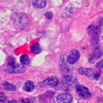
You just can’t put raw tissue or cell samples on your slides and expect good histology results! Instead you must preserve or ‘fix’ your samples. Fixing ensures that your cell structures stay intact and that your antigens are immobilized. Ideally, fixation would also still permit unfettered access of your antibodies to your antigens. However, as…