You’ve opened a plasmid map, but you have no idea what some of the features mean and how to work out whether this plasmid fits your experiment. And there’s so much to look at, including size, origin, markers, restriction sites, promoters, and tags. Where do you start?
This article walks you through the diagram feature by feature so you can look at any plasmid map and know what you’re talking about.
What is a Plasmid Map?
A plasmid map is a circular diagram of every detail of a plasmid, such as its size, the origin of replication, selectable markers, restriction sites, and any promoters, tags, and genes.
Read correctly, it tells you in under a minute whether a plasmid fits your experimental plan. But read carelessly, it sends you into a cloning or expression experiment that was never going to work! Therefore, the plasmid map is the first thing to check when a plasmid lands in your inbox, before you order primers, before you pick enzymes, before…anything!
Choose a free resource to help you move forward
DOWNLOAD
Nanodrop Results Interpretation Card
download
The Molecular Cloning Cheat Sheet
If you already know this plasmid is the wrong type for your experiment and need to pick a different one, check out our article on choosing the right plasmid vector instead.
Reading the plasmid map feature by feature
Every plasmid map is built from the same handful of features. Once you can name each one and know what to check, any map becomes readable. Including one you’ve never seen before! Here are the key features:
Size (the number in the center)
The center of the map gives the plasmid’s size in base pairs. pBR322, the classic example in Figure 1, is 4,361 bp when linearised. This is the number your diagnostic gel has to match.
Restriction sites (the labels around the outside)
The text around the outside of the map, often in blue, marks restriction sites: the positions where the plasmid is cut if you incubate it with the matching restriction enzyme (Figure 1). Each site is drawn as a short line at its starting nucleotide position.
Together, these sites make up the plasmid’s restriction map, the pattern of where each enzyme cuts (working that pattern out from digests is what’s meant by plasmid mapping). A site that appears once is a unique (single) cutter, and unique cutters in the multiple cloning site are what you use to open the vector cleanly. If you’re new to these enzymes, see 10 things you need to know about restriction enzymes.
Multiple cloning site (MCS)
The MCS is a short stretch, usually next to the promoter, packed with unique restriction sites. It’s where you insert your DNA. When you pick an enzyme from the MCS, run your insert sequence through the same enzyme first: if the enzyme also cuts your insert, it will fragment your gene instead of just opening the vector.
Origin of replication (ori)
The ori is the sequence where replication starts, and it sets the copy number. Don’t change it. Two plasmids that share the same ori are incompatible in a single cell, as one is lost over successive generations even if they carry different antibiotic resistance markers. If you’re co-transforming, this is the feature that decides whether both plasmids survive. The E. coli plasmid origins of replication series covers this in depth.
Selectable marker (antibiotic resistance)
The resistance gene lets you select cells that took up the plasmid. pBR322 (Figure 1) carries two transcribed in opposite directions: tet (tetracycline resistance) and amp (ampicillin resistance). Match the marker to an antibiotic you can actually use. Note that the β-lactamase encoded by amp is not selective between penicillin-derived antibiotics, so ampicillin resistance also confers resistance to related β-lactams.
Transcription direction (the arrows)
The arrows show the direction of transcription, which is essential for cloning (Figure 1). A gene has to sit downstream of, and in the same direction as, the promoter that’s meant to drive it. Get the orientation wrong, and a native promoter elsewhere on the plasmid can interfere with expression of your gene.
Promoter
The promoter drives transcription of the insert, and it must match your host. A T7 or tac promoter works in E. coli; CMV or EF-1α is for mammalian cells; GAL1, ADH1 or AOX1 for yeast; SP6, T7 or T3 for in vitro transcription. A plasmid with a bacterial promoter will not express your protein in mammalian cells, however healthy the map looks.

Terminator
The terminator stops transcription cleanly at the end of the gene. The pTLNX vector (Figure 2) pairs its SP6 and lacUV promoters with an rrnBT2 terminator for efficient termination. For in vitro transcription, the terminator (or the site where you linearise the plasmid) defines the 3′ end of your RNA.
Tags
Purification and detection tags (His, GST, GFP and others) are drawn next to the insert site and are labeled by the terminus they end up on, i.e., N-terminal or C-terminal of your translated protein. Which terminus matters: an N-terminal tag can block a signal sequence, and a C-terminal tag is only added if there’s no stop codon in between. Read the terminus off the map before you clone.
Reporter genes and primer binding sites
A reporter (GFP, lacZ) may be fused in frame to your insert as a readout of your protein, or sit in its own cassette as a transfection or screening marker. The map shows which promoter and reading frame run where. Primer binding sites (e.g., T7, SP6, M13) flank the MCS and are what you use to sequence across your insert.
A promoter that doesn’t match your host is not a minor detail — it’s the difference between a construct that expresses and one that never will, no matter how clean your cloning is.
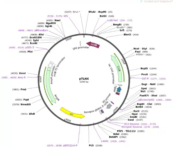
What the whole map is telling you
Reading features one at a time isn’t the point. What matters is what the plasmid map says as a whole. For expression, trace the map as a single unit: promoter → cloning site/insert → tag → terminator, all running in the same direction. If that chain is intact and the promoter matches your host, the plasmid can express your gene. If any link is missing or facing the wrong way, it can’t.
| MCS but no promoter near it | Cloning/storage vector — carries your gene, won’t express it | Whether you need expression at all; if so, move to an expression vector |
| Promoter → insert → terminator, same direction | A complete expression cassette | The promoter matches your host, and the tag is on the terminus you want |
| Two oris and two markers | Shuttle vector for two hosts | Which ori/marker pair applies to the host you’re using now |
| Phage promoter (SP6/T7/T3) beside the insert | In vitro transcription vector | The promoter matches your RNA polymerase; linearise downstream before transcribing |
When you present or share the plasmid, cite the repository accession (e.g. the Addgene number) rather than a redrawn map, so anyone can pull the verified sequence.
My plasmid looks wrong
Reading a plasmid map is only useful if it is accurate. These are the failures that trace back to the map, and what to do about each:
The linearised plasmid runs at the wrong size
Most likely cause: the tube isn’t the plasmid the label claims, or the map is out of date after an earlier cloning step.
What to do: run a diagnostic digest with two or three enzymes and compare the fragments to the map’s prediction. If they disagree, sequence before you go further. Don’t build on an unverified backbone.
My “unique” cutter cuts the plasmid more than once
Most likely cause: the site is unique in the vector but also present in your insert, or the map only shows sites in the MCS and hides duplicates elsewhere.
What to do: run both the full sequence and your insert through the enzyme. Pick a site that’s genuinely unique across the whole construct, or switch to a different enzyme or a partial digest.
Cloning worked, but the protein never expresses
Most likely cause: the promoter doesn’t match your host, or the insert went in facing away from the promoter.
What to do: confirm the promoter is right for your host and that the transcription arrow points toward your insert. Re-check the orientation with a directional diagnostic digest.
My tagged protein has no tag, or the tag kills its function
Most likely cause: the insert is out of frame with the tag, or the tag is on the terminus that blocks a signal sequence or active site.
What to do: read which terminus the tag sits on, confirm the insert is in frame with no intervening stop codon, and move the tag to the other terminus if it interferes.
One of two co-transformed plasmids keeps getting lost
Most likely cause: both plasmids share the same origin of replication, so they’re incompatible. Different resistance markers don’t rescue this.
What to do: check the ori on both maps. Use plasmids with different origins (different incompatibility groups) and keep both antibiotics in the medium. See understanding plasmid incompatibility.
What the map doesn’t tell you
- A plasmid map is a claim until you’ve cut and sequenced it. Maps are drawn from a reference sequence, and the tube in your freezer may have drifted from it through earlier cloning, a mislabel, or a mutation. Treat the map as a hypothesis you confirm with a diagnostic digest and a sequence read of the region you care about. This is why a repository map (Addgene) beats a homemade one: it’s tied to a deposited, verified sequence.
- Same origin of replication means incompatible and resistance markers don’t change that. Beginners assume two plasmids with different antibiotic resistances will happily coexist. They won’t if they share an ori. Replication competition slowly evicts one of them, and you end up selecting a population that quietly dropped a plasmid. When you plan any co-transformation, match ori to different incompatibility groups first, then worry about selection.
- Which terminus your tag lands on affects your protein. The map labels a tag as N- or C-terminal for a reason. An N-terminal tag can mask a signal peptide and mislocalize the protein; a C-terminal tag is only translated if nothing upstream introduces a stop. The map tells you the terminus; whether that terminus is safe for your particular protein is a judgment the map can’t make for you.
- Ampicillin selection fades in dense or old cultures. β-lactamase is secreted, and in a crowded or overgrown culture it degrades ampicillin in the medium, meaning untransformed cells start surviving too. For reliable selection, use fresh plates, don’t over-incubate, and consider carbenicillin for more stable selection. The map shows you the marker; it can’t warn you that the selection weakens over time.
Common mistakes
| Treating a cloning vector as an expression vector | No promoter upstream of the MCS on the map; protein never appears despite clean cloning | Confirm a host-matched promoter points at the insert before you commit to the vector |
| Choosing an MCS enzyme that also cuts the insert | Extra bands after digestion; the insert is fragmented, not just released | Run your insert sequence through the same enzyme; pick a site unique to the whole construct |
| Ignoring transcription direction | Construct verifies by size but doesn’t express, or expresses erratically | Follow the arrow: the insert must sit downstream of, and in line with, its promoter |
| Trusting a homemade or outdated map | Diagnostic digest fragments don’t match the map’s prediction | Use a repository (Addgene) map tied to a verified sequence; re-map after every cloning step |
| Co-transforming two same-ori plasmids | One plasmid is progressively lost despite double antibiotic selection | Check both oris; use different incompatibility groups for plasmids that must coexist |
| Reading a tag without checking its terminus | Tag missing on the purified protein, or the protein is mislocalised/inactive | Read the N/C terminus off the map; confirm the insert is in frame with no stop codon before the tag |
Originally written by Vicki Doronina. Renovated with feature-by-feature map reading, troubleshooting, and practitioner notes. Last reviewed July 2026.
References & further reading
More on working with plasmids and their features:
- Choosing the right plasmid vector — if the map tells you this isn’t the plasmid you need.
- Understanding plasmid incompatibility — why the ori decides whether two plasmids can share a cell.
- Three important things to check after obtaining your plasmid — the incoming-plasmid QC routine.
- E. coli plasmid origins of replication — the origin, copy number, and compatibility in depth.
- 10 things you need to know about restriction enzymes — for reading and choosing restriction sites.
- pUC18: probably the best high-copy plasmid in the world — a worked example of a common cloning vector.
Primary references:
- Watson N (1988) A new revision of the sequence of plasmid pBR322. Gene 70:399–403. DOI: 10.1016/0378-1119(86)90307-0
- Structural and functional characterization of plasmid features. Biochemistry. DOI: 10.1021/bi200178z
You made it to the end—nice work! If you’re the kind of scientist who likes figuring things out without wasting half a day on trial and error, you’ll love our newsletter. Get 3 quick reads a week, packed with hard-won lab wisdom. Join FREE here.