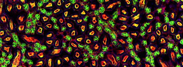
Confocal Laser Scanning Microscopy Explained In 3 Easy Steps
Learn how confocal laser scanning microscopy works, its applications, and why it’s great for samples that are too thin to section.
Join Us
Sign up for our feature-packed newsletter today to ensure you get the latest expert help and advice to level up your lab work.

Learn how confocal laser scanning microscopy works, its applications, and why it’s great for samples that are too thin to section.

Whether you want to get started with fluorescence microscopy or already use it, this guide will ensure you know the basics and get the best out of your fluorescence microscopy.

Learn how the Point Spread Function affects what you see through your microscope and discover what you can do to improve your images.

Discover a way to study the active form of proteases in situ with in situ zymography. Read this article to get up to speed.
When we are doing a double labelling, it makes good sense to choose two fluorophores with spectra widely spaced apart to avoid mixing. Why combine GFP with Rhodamine, when you can combine it with Texas Red?

Taking publication quality immunofluorescent images of can be a very time intensive, and frustrating process with hours spent capturing, processing, and putting the images into final figure format. And, if you aren’t careful, you can do a lot of work only to realize later that you need to re-image something for one reason or another….

The eBook with top tips from our Researcher community.