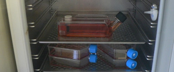
There is a growing trend in using microalgae as the expression system for heterologous proteins. However, I find most protocols dealing with microalgae available online are not that great or informative! So I would like to share my experience in using Chlamydomonas reinhardtii as the model microorganism.
Before you start expressing your protein, you need to confirm that your gene of interest (GOI) is in the microalgal genome. You can confirm this easily by genomic DNA extraction followed by colony PCR.
Genomic DNA Extraction
Similar to DNA isolation in other organisms, you need to understand the structure of the organism for efficient DNA recovery. Microalgae often have an intact or partially-disrupted cell wall, which poses difficulties in genomic DNA extraction. Therefore, a chelex resin is used in the DNA extraction procedure. Chelex stands for Chelating Ion Exchange Resin. Chelex is composed of styrene divinylbenzene copolymers containing paired iminodiacetate ions that act as chelating groups in binding polyvalent metal ions such as magnesium (Mg2+). Removal of Mg2+ from the reaction inactivates nucleases and protects the DNA. Chelex is not only used in a mixture, but you can also use it in a column-based format to purify proteins (depending on the application) and for whole blood DNA extraction from blood spotted on to a filter. It is also a very economical choice!
To extract the DNA follow the procedure from Berthold, Best, & Malkin1, which is summarized below:
- Pick a small amount of Chlamydomonas reinhardtii cells from each colony and dilute in 10µl double distilled water
- Add 10 µl of 100% ethanol and 100 µl of 5% (w/v) Chelex
- Heat the mixture at 99°C for 5 minutes (Optional: place on ice for an additional 5 min)
- Centrifuge at 13,000 rpm for 5 minutes
- Use 1µl of the supernatant containing the isolated genomic DNA for PCR
- Store the rest of the supernatant at -20 °C for later use
Colony PCR
The conditions for colony PCR really depend upon your primers and GOI, so I can’t say much about that! But I can offer some general troubleshooting tips that may be useful.
- Use a minimal amount of cells (you should just see a very faint green color in the whole mixture), otherwise it’ll be too dense and inhibit the PCR reaction
- Always use healthy cells on plates, otherwise you won’t get clear bands after gel electrophoresis. And all of the previous work is in vain!
- Always have a positive and negative control (with only water!) during PCR
- Try to load the samples at the bottom of the PCR tube without touching the side of the tube
- Always add the polymerase last and the component with the largest volume (it’s water in my case) first
- Always close the PCR tube cap properly, especially when you use PCR strips! (I once lost most of my samples due to evaporation because I didn’t close the caps completely.)
All the best with everyone’s lab work!
Reference
- Berthold DA, Best BA, Malkin R. (1993) A rapid DNA preparation for PCR from Chlamydomonas reinhardtii and Arabidopsis thaliana. Plant Mol Biol Rep. 11(4):338-44.