Think of the very first time you looked at cells under a table-top microscope. Here’s what you would have done in that experiment:
Step 1: Grow cells.
Step 2: Plate cells on to a glass/quartz slide.
Step 3: Insert the slide under a microscope and look.
Enjoying this article? Get hard-won lab wisdom like this delivered to your inbox 3x a week.

Join over 65,000 fellow researchers saving time, reducing stress, and seeing their experiments succeed. Unsubscribe anytime.
Next issue goes out tomorrow; don’t miss it.
The protocol for performing single-cell microscopy has a similar skeleton. However, things get detailed when you want to obtain single-cell resolution and ensure that your data is biologically relevant and reproducible.
Why single cells?
Over the last few decades, we have realized that no two cells are absolutely identical, even those that contain the same DNA. Individual cells that receive the same signal can respond differently. Such diversity in cellular response is widespread among all cell types and most biological processes. This is particularly true when performing cancer cell microscopy, where you may want to understand the heterogeneity in the cancer sample.
To completely understand how cells interact to form living tissue, organs and organisms, it is crucial to observe how individual cells behave.
This is where single-cell microscopy comes into play.
Using this approach, you can watch large numbers of individual cells in real-time and figure out how they communicate with each other and respond to environmental cues.
Here, I describe three aspects of the experimental setup and design in single-cell microscopy that you need to consider.
Time-lapse imaging
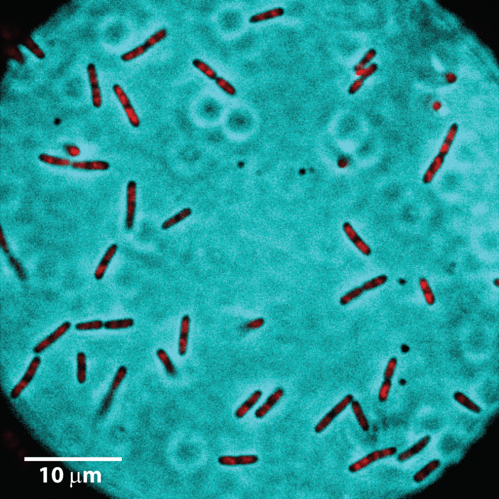
Although some experiments might involve static, one-time snapshots of single cells (see Figure 1); these provide an incomplete picture of the cellular processes. Cells are not mute spectators to the outside world! They are dynamic and continuously modulate their response through internal signaling pathways, so it is important to study their behavior over time. In other words, you would want to shoot a time-lapse movie of the cell to see how it responds in your experiment. And once the experiment is over, the movie can be saved and played back for in-depth analysis (see video below).
This time-lapse movie shows single E. coli cells dividing to form daughter cells. Total imaging duration is 59 min. (Data courtesy Weisshaar Lab, University of Wisconsin-Madison)
You will need to watch the same group of single cells over a period of time. To achieve this, you need to maintain a subset of cells in focus of the microscope objective. This is where commercially available temperature controllers and perfect focus systems come in use.
You can also automate instrument handling (shutters for light sources, camera acquisition sequences, filter wheels, and solution flow) and image analysis using open-source software packages such as Micro-Manager and Fiji. You don’t have to be a programming whiz-kid to do this – anyone with interest in writing small bits of code can learn the ropes, especially with the help available in online forums.
Flow chambers
You need a flow chamber to house the cells during the experiment. This usually consists of an enclosed volume with the top or bottom surface made of a microscope glass coverslip, and equipped with inlet and outlet ports through which you can flow growth medium to keep cells happy. The chamber rests on or under the microscope objective (for inverted or upright microscopes, respectively) placing the cells within viewing distance. It is common to maintain the chamber and, therefore, the cells, at a constant temperature using heating cables.
Nowadays, microfluidic chambers have become quite popular for trapping single cells. These have orders-of-magnitude smaller volumes than macroscopic chambers and enable lower consumption of resources per experiment.
Are the cells alive?
Depending on the experimental design, you may or may not want the cells to be alive and ‘happy’. If you want to do live cell imaging, then you need to ensure that you are supplying enough nutrients (oxygen, carbon source, amino acids, nucleotides) and growth medium at the right pH, temperature, and osmolarity. Constant flow of growth medium and temperature control of the flow chamber can help you achieve this.
If you are studying fixed cells, be aware that the fixation process itself can cause major artifacts in the observations. For example, protein localization can be affected a great deal!
Cellular adhesion to the glass surface is likely to affect growth rate. However, this might be within tolerable limits for the experiment. However, it is a good idea to check that the slide coating agent (e.g. poly-Lysine for bacteria, Concanavalin A for yeast) does not cause artifacts in the setup.
Finally, if you are imaging fluorescent reporter molecules using a high-power laser, check for laser-induced cell damage. Certain imaging methods require use of high laser powers (>1 kW.cm-2), which can cause irreversible damage to the cell within a few imaging cycles. Specifically, check cell viability and growth rate; and compare it with behavior of cells growing in culture tubes.
Watching single cells in real time is a powerful approach to understanding cellular function. It bridges the gap between research and those amazingly fabulous images that you see on science TV, and is also a great way to spend your workday!
You made it to the end—nice work! If you’re the kind of scientist who likes figuring things out without wasting half a day on trial and error, you’ll love our newsletter. Get 3 quick reads a week, packed with hard-won lab wisdom. Join FREE here.