Confocal Laser Scanning Microscopy Explained In 3 Easy Steps
Learn how confocal laser scanning microscopy works, its applications, and why it’s great for samples that are too thin to section.
Join Us
Sign up for our feature-packed newsletter today to ensure you get the latest expert help and advice to level up your lab work.
Learn how confocal laser scanning microscopy works, its applications, and why it’s great for samples that are too thin to section.
Not sure what FRET is, or just need a refresher on how FRET works? Read our short guide to understand the usefulness of FRET for studying protein-protein interactions.
There are those of us who began our careers literally in the dark. Yes, there was a time and not that long ago, that all figures had to be on film. Slide presentations were slides. Micrographs were, well, micrographs on film. Figure creation involved several steps: figures for publications had to be mocked up; then…
We can, however, use special dyes or engineered proteins to bind to calcium, indicating where in the cell it is. The trick is determining which probe is most suitable to your cells and application.
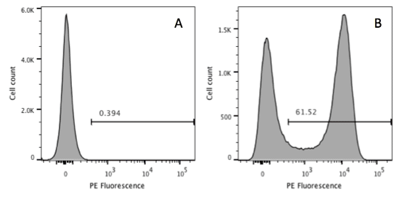
In many biological experiments the question that a researcher wants to ask is – ‘do some or all of my cells express a particular protein?’ There are many ways of doing this, which you will be familiar with e.g. Western blotting, immunoprecipitation, microscopic examination of stained cells and even mass spectrometry. Using Flow Cytometry to…
If you want to see in real time what is going on inside your cell then you should be performing live-cell imaging. Live-cell imaging techniques allow real-time examination of almost every aspect of cellular function under normal and experimental conditions. With all live-cell imaging experiments, the main challenges are to keep your cells alive and healthy…