Ultramicrotomy:
The Cutting Edge of Sectioning

Viola Oorschot
Technical Officer, Electron Microscopy Core Facility at EMBL Heidelberg
Read BioViola is responsible for running facility EM projects at the Electron Microscopy Core Facility at EMBL using techniques including cryo-immuno EM, micro CT scan, and recently 3D volume EM. She has previously worked at Monash University Melbourne and University Medical Centrum Utrecht, working with Judith Klumperman, Hans Geuze, and Jan-Willem Slot. Her experience includes Tokuyasu EM and Correlative Light and Electron Microscopy (CLEM) techniques for cell biology and biomaterials, as well as X-ray target CLEM, SEM, and STEM.
Close
Jemima Burden
Head of Electron Microscopy, MRC Laboratory for Molecular Cell Biology, University College London
Read BioJemima is Head of Electron Microscopy at the Laboratory for Molecular Cell Biology at University College London. She received her BSc from the University of Bath and her PhD from Imperial College London, where her fascination for microscopy began. Her main interests are developing Correlative Light and Electron Microscopy (CLEM) workflows and using volume EM to investigate cell biological processes from viruses to whole organisms.
Close
José Maria Mateos Melero
Research Associate, Center for Microscopy and Image Analysis, University of Zurich
Read BioJosé María obtained his PhD in Neuroscience at the Basque Country University. He worked as a postdoc at the Brain Research Institute and Institute of Biochemistry of the University of Zurich. Currently, he is a Research Associate at the Center for Microscopy and Image Analysis of the University of Zurich.
Close
Nicolas Schilling
Research Associate, Center for Microscopy and Image Analysis, University of Zurich
Read BioNicolas obtained his Master in Biomedicine with a focus on biomedical imaging at the Center for Microscopy and Image Analysis of the University of Zurich where he is currently working as Research Associate.
CloseDiscover ways to refine your ultramicrotomy technique to improve electron microscopy (EM) imaging for a wide range of samples and EM techniques.
In this webinar, you will learn:
- Ways experts refine their ultramicrotomy workflows for specific applications such as volume EM.
- How to use more targeted sample sectioning for advanced imaging.
- Methods for dealing with samples that demand array tomography or serial sectioning.
Learn how to do more with your samples, thanks to better sample preparation! The ability to image biological ultrastructure in EM relies on the sample preparation quality, but improving your workflow doesn’t necessarily mean a new setup is required. You’ll be surprised what improvements you can make by simply unlocking the full potential of your ultramicrotomy workflow.
Join this webinar where experts from three world-renowned institutions at the cutting edge of life science research share the latest advances in techniques in their workflows for a wide range of samples, with techniques such as volume EM, Tokayasu-CLEM, and array tomography.
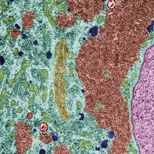
Ultramicrotomy is a proven and universally accepted sample preparation technique for TEM, SEM, AFM, and more. It permits visualization and analysis of the fine internal structures of samples at nanometer-scale resolution by producing ultrathin sample sections quickly and cleanly. Ultramicrotomy offers several key advantages, including the size and homogeneity of the electron-transparent area within the sections and the speed of section production.
AGENDA
Welcome and introductions
Target trimming of resin blocks with crosshair for volume EM
Viola Oorschot, EMBL
Sectioning for array tomography – a quick start guide
Jemima Burden, UCL
Ultramicrotomy for diverse projects in an imaging facility: methods including 2D sections, Tokuyasu-CLEM, and array tomography
José Maria Mateos Melero & Nicolas Schilling, University of Zurich
Q&A session and panel discussion