Revealing Cellular Dynamics with Millisecond Precision – The New Tool That Turned Electron Micrographs into Motion Pictures of Neural Communication
Dr. Shigeki Watanabe
Associate professor of cell biology, Johns Hopkins School of Medicine, Solomon H. Snyder Department of Neuroscience
Read BioAbout you (short Bio, 450 characters maximum): Shigeki Watanabe was trained in a broad range of techniques for electron microscopy. He has applied morphological analyses to study neurotransmission in worms and mice. He has developed two novel approaches in electron microscopy: protein localization by super-resolution microscopy and time-resolved analysis using optogenetics. He received his PhD from University of Utah in 2013. He completed his post-doctoral trainings in the laboratory of Erik Jorgensen and Christian Rosenmund. He is now an Assistant Professor at the Johns Hopkins School of Medicine
Close
Dr. Frédéric Leroux
Advanced workflow specialist, Leica Microsystems
Read BioFrédéric Leroux completed his Master degree in Biology in 2007 at the University of Ghent where he gained experience in biological EM sample preparation. In 2008, he moved to the physics department at the University of Antwerp where he obtained his PhD in 2012. At the EMAT research group he specialized in advanced electron microscopy of composite materials and polymers. In 2016, he joined Leica Microsystems as Application Specialist Nanotechnology. He thereby uses his multidisciplinary background and broad microscopy experience to improve EM sample preparation of a variety of materials (polymers, composites, biological and industrial materials).
CloseIn this tutorial:
- What if you can dissect the cellular dynamics with millisecond precision?
- What if you can unravel the morphological transformation of a neuron millisecond by millisecond using electron microscopy?
- Could this be even possible?
In this webinar, we will talk about how optogenetics coupled with high-pressure freezing can make this possible.
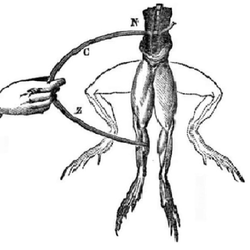
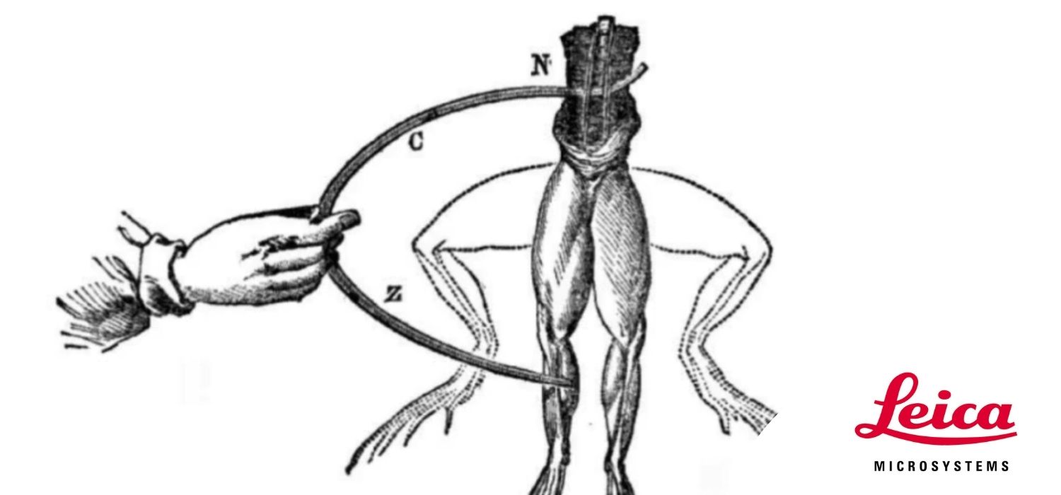
We will discuss how to implement electrical stimulation and why it is superior to light stimulation.
We will also discuss the importance of sample processing and the challenges you would face while freezing different types of samples.
View the webinar replay to hear the story of discovery by our speakers Dr. Watanabe and Dr. Leroux.