Hyperplex Cancer Tissue Analysis at Single Cell Level With Cell DIVE™

Nicolas Mouchet, PhD
Platform Manager H2P2 BIOSIT Université de Rennes
Read BioNicolas Mouchet has been co-manager of the H2P2 histopathology platform at the University of Rennes since 2019. He completed his engineering degree in 2006, gaining experience in molecular genetics and RNA expression analysis. During his PhD and post-doctoral studies, he trained in cancer research bioinformatic analysis. In 2017, he earned a Master’s degree in Computer Science to better support the evolving cross-disciplinary nature of scientific studies.
Close
David Pointu, PhD
Senior Application Manager Cell DIVE and Translational Research Leica Microsystems
Read BioDavid Pointu has been application manager for Cell DIVE and translational research at Leica Microsystems since 2021 and has broad experience in light microscopy, including various aspects of cell and tissue imaging and analysis. After studying physical chemistry at the University of Strasbourg, he completed his PhD in 2002, followed by a postdoctoral fellowship at the Institut Curie. In 2004 he moved into industry, and has worked as an application specialist in high-end microscopy at several companies, including GE Healthcare.
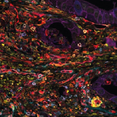
CloseThe H2P2 histopathology platform team have been mastering immunohistochemical staining and multiplexing for more than 15 years. With Cell DIVE technology, they offer the scientific community the opportunity to perform hyperplex labeling with up to 50 labels on a single tissue section. Simplicity of implementation, combined with an original acquisition system, makes this technology especially valuable in immuno-oncology research.
Cell DIVE technology allows researchers to investigate crucial topics such as lymphoma cell heterogeneity and the nature of relapses and therapeutic resistance. How lymphoma cell heterogeneity influences the response to their microenvironment can be studied at mutational, transcriptomic, and protein levels. However, because protein expression studies provide the most relevant information about the nature of cellular interactions and protein expression levels, Cell DIVE makes it possible to address these topics in the context of immunological synapse spatial organization.
In this webinar, you will discover:
- How the hyperplex workflow can be applied for studying multiple proteins from the same cancer tissue;
- More about the technical aspects of the hyperplex workflow;
- How Cell DIVE technology enables spatial profiling;
- Why image analysis in immuno-oncology is important.