Exploring The Structure and Life Cycle of Viruses The Virologist’s Toolbox for Electron Microscopy Specimen Preparation
Frédéric Leroux
Advanced Workflow Specialist Leica Microsystems
Read BioFrédéric Leroux completed his Master degree in Biology in 2007 at the University of Ghent where he gained experience in biological EM sample preparation. In 2008, he moved to the physics department at the University of Antwerp where he obtained his PhD in 2012. At the EMAT research group he specialized in advanced electron microscopy of composite materials and polymers. In 2016, he joined Leica Microsystems as Application Specialist Nanotechnology. He thereby uses his multidisciplinary background and broad microscopy experience to improve EM sample preparation of a variety of materials (polymers, composites, biological and industrial materials).
CloseIn this webinar, you will learn about:
- Specimen preparation workflows for studying the viral structure and life cycle
- Room temperature, hybrid and cryo workflows
- Correlative microscopy
- Technical aspects of the workflows
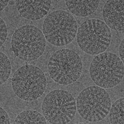
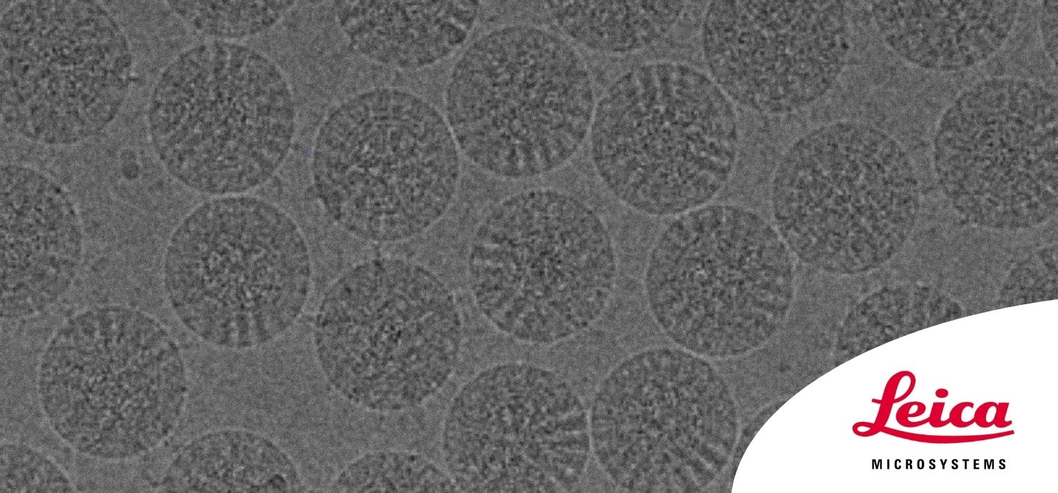
The SARS-CoV-2 outbreak started in late December 2019 and has since reached a global pandemic, leading to a worldwide battle against COVID-19. Understanding the structural components of viruses, host-interactions, and their intracellular life cycle will support and accelerate vaccine and anti-viral agent design. Because of its resolution, electron microscopy is a powerful and essential tool in the field of Virology.
The ever-evolving electron microscopy methods offer a plethora of new applications, allowing researchers to study viral structure, infection and replication. During this webinar, we provide an overview of these sophisticated techniques and illustrate how they provide insights into the intricate changes that viruses evoke when they infect cells. We cover the currently used preparation workflows for SEM and TEM, including room temperature, hybrid and dedicated cryo workflows.