Zeiss
A Beginner’s Guide to The Point Spread Function
Learn how the Point Spread Function affects what you see through your microscope and discover what you can do to improve your images.
Read MoreAn introduction to Photoactivated Localization Microscopy (PALM)

How does photoactivated localization microscopy (PALM) work? And what use can PALM microscopy be to you? This short introduction to PALM gives you the answers!
Read MoreBeginners Guide to PALM Sample Preparation

Good PALM sample preparation is the key to great images. Find out how to choose the right fluorescent proteins and learn some tips and tricks for sample prep.
Read MoreAutomated Microscopy
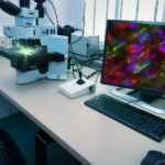
The traditional microscope that you know and love is operated manually. Picture the scene: the microscopist chooses the light source, gently places the sample the moveable stage, selects the objective lens, and scans to select the field of view. This process is perfect for processing and analyzing a small number of samples per day. But…
Read MoreData Analysis for Three-dimensional Volume Scanning Electron Microscopy
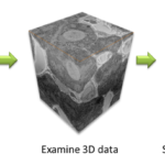
In recent years, three-dimensional (3D) scanning electron microscopy techniques have gained recognition in the biological sciences. In particular, array tomography, serial block face scanning electron microscopy (SBFSEM) and focused ion beam scanning electron microscopy (FIBSEM) (described in Three-Dimensional Scanning Electron Microscopy for Biology) have shown an increase in biological applications, elucidating ultrastructural details of cells…
Read MoreThree-Dimensional Scanning Electron Microscopy for Biology
Scanning electron microscopy (SEM) is a powerful technique, traditionally used for imaging the surface of cells, tissues and whole multicellular organisms (see An Introduction to Electron Microscopy for Biologists)(Fig. 1). While the resultant images appear to be three dimensional (3D), they actually contain no depth information. However, there are several SEM techniques that can obtain…
Read MoreLet There Be Light! Microscope Maintenance Part 2: Köhler Illumination

In Part 1 of these articles, you’ll have learnt about common microscope light sources and how to replace and align these correctly. In this article, we will discuss the importance of Köhler illumination and how to set up the microscope to achieve optimal imaging results. What is Köhler illumination? Before discussing this technique, let us…
Read MoreLet There Be Light! Microscope Maintenance Part 1: Routine Care and Replacing Bulbs

Do you want the best imaging experience each time you use a microscope? Well, this is a rhetorical question, as we all desire that these delicate optical instruments are clean, free from immersion oil and correctly aligned. From the routine checking of slides, capturing images for presentations and publications, to diagnosing diseases using point-of-care microscopes,…
Read MoreHow to Troubleshoot Problems with Fluorescently Tagged Proteins
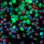
Using fluorescent proteins as imaging probes is a widespread and versatile technique in microscopy. You can use them in a wide range of living systems, from single cultured cells to complete organisms and animals. Fluorescently tagged proteins can be used to track and examine real-time localization, interactions and translocation of your protein of interest, as…
Read MoreLive-Cell Imaging: Choosing the Right Technique
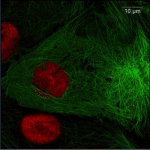
If you want to see in real time what is going on inside your cell then you should be performing live-cell imaging. Live-cell imaging techniques allow real-time examination of almost every aspect of cellular function under normal and experimental conditions. With all live-cell imaging experiments, the main challenges are to keep your cells alive and healthy…
Read More