Crash Course in Microbial Identification
Need a crash course in methods for identifying microbes? Well, look no further: here, you’ll find an overview of the methods available for the identification of bacteria, yeast, or filamentous fungi to the species level.
Identifying Microbes at the Species level: The Why
Species-level identification allows you to discriminate between two species from the same genus, which is often essential in the treatment of infectious diseases. For example, the bacterial genus Yersinia contains approximately 15 species, some of which form part of the normal human microflora, but others of which are serious pathogens and require medical treatment (e.g. Yersinia pestis is the causative agent of bubonic plague).
Before we get into identification, let’s outline some of the main applications of accurate microbial identification:
- Healthcare – accurate and fast identification of bacteria, fungi, and parasites is important for correct and timely disease diagnosis, and appropriate treatment.
- Epidemiology – identifying microbes is important for tracking and tracing disease spread and outbreaks, as well as for identifying new isolates, e.g. antibiotic-resistant isolates.
- Pharmaceutical industry – because microbes are a significant threat to sterility, being able to accurately identify microbes is often a good manufacturing practice (GMP) requirement within the pharmaceutical industry.
- Additional scenarios – criminal investigations, microbial forensics (the investigation of bioterrorism threats), and environmental studies all need to accurately identify microbes for various purposes.
So, how do we go about identifying microbes? Traditional methods rely on phenotypic identification using staining, culturing, and simple biochemical tests. Nowadays, more powerful molecular, immunological, and biochemical analytical methods complement and sometimes replace traditional methods. We’ll look at both traditional and modern methods for identifying microbes below.
Traditional Methods for Identifying Microbes
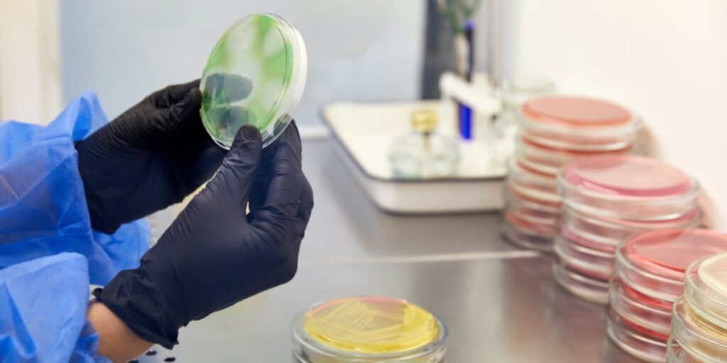
1. Macroscopic Features
Macroscopic features encompass the overall appearance of a microorganism, including its shape, size, color, and smell (i.e. the features that you can see with the naked eye). You can often determine the type of microorganism by examining the gross morphological/macroscopic features on an agar culture.
Examining Agar Cultures
- Filamentous fungi or molds appear as ‘hairy’ irregularly shaped colonies and often produce visible spores that may look powdery or dusty. Fungal colonies can contain more than one color, usually with a darker color in the (often raised) center and a lighter color radiating from it. Filamentous fungi grow radially from the center of an agar plate, with the youngest on the outside, and the older darker material (rich with spores) on the inside. Filamentous fungi may also grow as a unicolored furry mat with no obvious sign of spores at all!
- Bacteria often form distinct colonies, which are sometimes smaller than fungal colonies and can be anything from slimy to very dry in texture. They range in color from white to bright red.
- Bacteria often have a strong odor while filamentous fungi can be odorless or have an earthy smell.
- Yeast can be the trickiest microbe to identify based on macroscopic features, because colonies often look similar to bacterial colonies, depending on the species and type of agar used.
- The same species can appear differently depending on the culture medium.
2. Microscopic Features
The following questions can take you a long way in identifying microbes using the microscope:
- Are you looking at rods, cocci, or spiral-shaped bacteria?
- Are you looking at bacterial cells with flagella?
- Are you looking at budding yeast?
- Are you looking at a filamentous fungus with branching hyphae?
3. Staining and Microscopy
Stains enable easier visualization under a microscope. Cytology microscopes have specific requirements to ensure clear differentiation between stained cells. Here’s an overview of the most popular microbiological stains.
Gram Staining
Gram staining is often the go-to test in bacterial identification. This purple stain, based on the crystal violet dye, is named after the Danish bacteriologist Hans Christian Gram, who developed it.
Typical Gram-positive bacteria include Bacillus, Staphylococcus, Streptococcus, and Clostridium spp., while Escherichia, Helicobacter, and Salmonella spp. are Gram-negative. Certain bacteria are Gram-variable and, therefore, aren’t amenable to Gram-staining.
Endospore Staining
This involves applying a stain to a bacterial sample to check for the presence of spores. Because not all bacteria produce spores, this information can be useful in identification. Several spore stains are available, but malachite green is probably the most popular.
Ziehl-Neelsen Staining
This is an important tool for the staining of Mycobacterium tuberculosis (TB), which can’t be Gram-stained. The red stain carbol fuchsin is used first, followed by a counterstain such as methylene blue. M. tuberculosis stain red while other bacteria stain blue.
Stains for Fungi and Yeast
Several fungal stains exist, although they are generally non-specific. They help visualize fungal elements for identification rather than discriminate between fungal species. Examples of fungal stains include:
- Lactophenol cottonblue – this stains the carbohydrates in fungal cell walls blue.
- Periodic-acid Schiff stain (PAS stain) – this stains carbohydrates and other moieties and causes a magenta color in living fungi only. This is used in infection diagnosis.
- Grocott’s methenamine silver stain – this colors fungal cell walls brown to black, but cannot distinguish between live or dead material.
- Trypan blue, aniline blue, and calcofluor white – these stain fungal and plant structures and are used in the study of plant/fungal symbiosis and plant pathology.
4. Simple Biochemical Tests
Catalase Testing
- If the unidentified bacterial species has catalase activity, bubbles of oxygen appear when hydrogen peroxide is added to a bacteria scraping on a microscope slide.
- Staphylococci, Micrococci, E. coli, and the other Enterobacteriaceae, as well as Salmonella spp., are among the most routinely encountered catalase-positive bacteria, while Streptococcus and Enterococcus bacteria are catalase-negative.
Oxidase Testing
- The oxidase test identifies bacteria with cytochrome c oxidase (CCO) activity. This enzyme forms part of the bacterial electron transport chain.
- When present, CCO oxidizes the reagent (tetramethyl-p-phenylenediamine) to a purple-colored product.
- When the enzyme is not present, the reagent remains in the reduced state and is colorless.
- Although oxidase-positive bacteria are aerobic, they are not necessarily strict aerobes and may be capable of anaerobic respiration.
- You will obtain a false-negative result if your bacterial species possess an oxidase incapable of reacting with the test reagent.
Substrate Utilization Tests
- A range of substrate utilization tests for microbial identification is available commercially.
- These tests (usually for bacterial species) contain a panel of substrates, e.g. different carbon and nitrogen sources.
- Bacterial species use these substrates differentially, and a record of the color changes in substrates after incubation with bacteria generates a key (or pattern) of substrate utilization.
- This key is compared with the substrate utilization patterns of a computerized list of bacterial species.
- These tests are probably more reliable in combination with catalase/oxidase testing and microscopic examination.
These tests overlap with the concept of selective growth media. A wide range of selective media exists for the isolation and identification of bacterial and fungal species.
Physiological Requirements for Growth
Addressing the following questions can help you when identifying microbes:
- Does the organism require oxygen?
- In what temperature range does the organism grow best?
- Can the organism tolerate acidic media?
5. Dichotomous Identification Keys
Dichotomous keys contain a series of steps, with each step presenting descriptions of two distinguishing features (e.g. Gram-positive or Gram-negative), with a direction to the next step in the key, until the identity is known.
The idea is that you use dichotomous identification keys alongside the methods outlined above to help you identify your organism of interest.
Modern Methods for Identifying Microbes
Although still widely used, traditional methods for identifying microbes suffer from two major drawbacks. First, they are applicable only to organisms that can be cultured in vitro. Second, some strains exhibit unique biochemical characteristics that don’t fit the pattern of any known genus and species.
Fortunately, many modern methods for identifying microbes aren’t dependent on live cultures, and they can often reveal minute differences between organisms that escape detection by traditional means.
6. Identifying Microbes Using PCR
- PCR, including Real-Time PCR, is probably the most widely used molecular technique for identifying microbes. Using PCR, one can rapidly detect and identify microbial species directly from clinical samples, thus speeding up diagnostic procedures.
- Several PCR-based methods exist. Most involve a universal set of PCR primers that identify bacterial/fungal samples by sequencing the PCR amplicons.
- The 16S rRNA gene is the gold-standard sequence for bacterial identification by PCR, while the Internal Transcribed Spacer (ITS) region is the primary barcode marker for fungal species.
7. Microarray-Based Identification
- Microarray-based microbial identification relies on the hybridization of pre-amplified microbial DNA sequences to arrayed species-specific oligonucleotide probes. Each probe contains a distinct dye that fluoresces on hybridization.
- Microarrays are a versatile tool, facilitating the detection and discrimination of different microbial samples on a single slide. Microarray is a fast technique, and speed is important in clinical settings for diagnosis and the timely initiation of proper antimicrobial therapy.
- Microarray-based platforms typically use similar barcode regions as other PCR-based methods (above).
8. Immunological Identification
- ELISA-based methods can be set up for microbial detection (usually within diagnostics) on a species-by-species basis.
- These methods are highly sensitive but they rely on very specific antibodies and highly discriminating protein(s) within the organism of interest.
9. Chemical/Analytical Identification
Fatty Acid Profiling
- Fatty acids are essential within bacterial cell membranes, and different bacterial species produce different combinations of fatty acids. This fatty acids profile can be used to determine an unidentified bacterial species by matching against known profiles.
- Fatty acid profiling is typically performed using a combination of gas chromatography and mass spectrometry.
Metabolic Profiles/Chemo-Profiling
- Besides primary metabolites (e.g. ATP, ADP) that are essential for growth, microbes produce a plethora of secondary metabolites that aren’t essential for growth but may be advantageous within certain environments, e.g. outcompeting other microorganisms for nutrients.
- Secondary metabolites include antibiotics, immunosuppressive compounds, pigments, and antioxidants. These metabolites are a huge source of existing and newly emerging drugs.
- Different species often produce unique secondary metabolite profiles, which allow us to identify known microbial species. Metabolic profiling is usually carried out using HPLC and mass spectrometry.
Do you use any other methods for identifying microbes? Just drop us a line in the comments section and let us know!
Resources
- Printable dichotomous identification keys for bacteria.
- Houpikian P and Raoult D (2002) Traditional and Molecular Techniques for the Study of Emerging Bacterial Diseases: One Laboratory’s Perspective. Emerg Infect Dis. 8(2): 122–31. doi: 10.3201/eid0802.010141.
- Järvinen A-K, Laakso S, Piiparinen P, Aittakorpi A, Lindfors M, Huopaniemi L, et al. (2009) Rapid identification of bacterial pathogens using a PCR- and microarray-based assay. BMC Microbiol. 9:161. doi: 10.1186/1471-2180-9-161.
- Khot Prasanna D and Fredricks DN (2009) PCR-based diagnosis of human fungal infections. Expert Rev Anti Infect Ther. 7(10):1201–21. doi: 10.1586/eri.09.104.
Originally published July 27, 2017. Reviewed and updated February 2021.