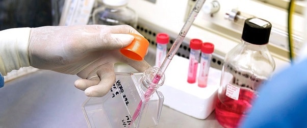
An Out of Body Experience: ex vivo Tissue Culture for Cancer Drug Screening

When choosing a model system for culturing tumor cells, we often think of the obvious tried and tested models. In vitro methods include cell lines that have been specifically selected to grow in culture flasks in incubators. While conveniently available, consistent and reproducible, cell lines are limited in that they may not represent the desired stage of disease or population heterogeneity. In vivo models require whole living organisms, for example, tumor tissues xenografted into mice. This method has the edge when testing the overall effects of a drug on a living subject, but require additional care and ethical approval.
Is there a model that combines the relative convenience of a cell culture system with the closer-to-clinical-conditions of a living system?
The answer may lie in the technique of ex vivo, or “out of the living”, tissue culture, which has undergone considerable resurgence recently with improvements in culture techniques and the need for a rapid method of shuttling cancer drugs from bench to bedside.
Why ex vivo?
Success rates for cancer drug clinical development are dismally low, with only 5% of anti-cancer compounds getting past phase III clinical trials. The translation of discovery to bedside can also be a prohibitively long and costly processes. Ex vivo culture is an efficient, accurate and clinically relevant pre-clinical model to understand the efficacy and mechanisms of action of new cancer drugs at a tissue level.
Ex vivo culture allows experiments to be performed on tissue (either intact, or cut into small pieces called an explant) in a dynamic but controlled environment. Experiments are performed under sterile culture conditions, generally following the drug-treated tissues for 24-72 hours.
Some obvious benefits include:
- The ability to maintain native tissue architecture (including the relationship between malignant cells and their surrounding stroma).
- Hormone responsiveness,
- Stromal-epithelial signaling
- Patient heterogeneity.
In the context of cancer drug screening, these characteristics allow differences to be seen between patients’ responses to the drugs, which has translational relevance. There is also the possibility of generating matching patient derived xenografts and cell lines for future experimentation, conceivably leading to precision medicine for that patient.
Which Method Should I Use?
The type of culture technique chosen depends on the type of tissue, length of treatment, and type of treatment. Three of the main options are the submersion, grid and sponge methods.
Submersion
Major limitations of the submersion method are that gentle rotation is needed to ensure proper oxygen availability to the sample, and that epithelial cells migrate to the cut surface of the tissue and produce a surrounding capsule.
Grids
The grid method is an adaptation of the submersion method which is used to minimize unwanted cell migration by supporting the tissue on a scaffold (e.g. lens paper or agar rafts) held up by a grid such as titanium. This method requires very thin slices (200-300 mM) and helps maintain cell viability for at least a week.
Sponge
Another scaffold that has recently shown promise is the gelatin or collagen sponge, which when soaked in drug-containing media acts as a nutrient/waste exchange site for the 1-2 mm3 tissue pieces resting atop it. Cell viability can be maintained and experiments of up to a week may be undertaken using this method.
Whatever the method chosen, these experiments should all demonstrate epithelial cell proliferation after 24 hours in culture, provided that the tissue pieces are sufficiently small enough for nutrient and oxygen perfusion.
Experimental Ooverview- How Does it Work?
Immediately following surgery, tissue cores from the organ or tumor are taken and dissected under sterile tissue culture conditions. These patient explants are placed into their culture dishes containing media and the drug of choice, supported by their scaffold or sponge. Samples are incubated under standard cell culture conditions for 2-4 days in most cases. At the end of the treatment, tissue pieces for each drug or treatment concentration are either fixed and embedded for immunohistochemical staining, snap frozen for RNA isolation and gene expression analysis, or made into protein lysates for western blot analysis.
Any Drawbacks?
- One obvious limitation of this procedure is the need for an ongoing source of fresh tissue specimens of the cancer of interest, which requires not only strong collaboration with surgeons and pathologists but also ongoing ethical approval.
- While the tumor tissues maintain the local features of the tumor, they no longer have functional vasculature nor a system of signaling with other organs and systems in the body.
- It will often be the case that the tumor being resected is from an early stage, treatment naïve cancer, which may exhibit different drug responses than a metastatic and drug resistant tumor would during later stages of cancer. This could lead to bias in the drug testing results.
Ex vivo culture as an environment for cancer therapy testing provides several unique facets to the pre-clinical testing required for patient efficacy. Because the method is patient specific, it may even have potential applications for precision medicine, including drug resistance testing, biomarker discovery, and dose optimization. Hopefully the rapid screening of the most promising agents flagged by this method will expedite the drug discovery to bedside pipeline.