Confocal Images: Expert Tips for Your Hour of Need!
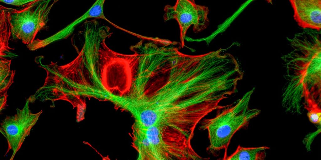
Are your confocal images leaving you with something to be desired? Perhaps you think your images are good, but you’re wondering if they can be better? I’ve put together some top tips to get your confocal images looking the best they can.
1. Selecting Your X/Y Resolution
There are other great articles where you can read about resolution in a confocal microscope in more detail, but to make a long story short, to get proper digitization of your signal, your digitization resolution must be at least twice the resolution you are trying to achieve. In other words, you must pick a sampling rate (pixel density) that gives you twice the resolution of your lens.
Confocal microscope software helps you do this. By giving you the details of your lenses, and what resolution you will achieve with what sampling (i.e. 1024 x 1024 or 4096 x 4096 pixels). You should choose to sample at a pixel rate that gives you twice the resolution (half the micron) that the specific lens gives you. Some microscopes make you really lazy and even have an “optimize resolution” button. These buttons take into account the lens, the wavelength, the pinhole, and the zoom factor to set the pixel ratio. And all you have to do is click it.
2. Selecting Your Z-step Resolution
Similar considerations apply to z-resolution. Of course, optical resolution in z is much less to begin with, so you will end up needing fewer pixels. If you want to do 3-D reconstruction, z-projection, or morphometry, you should always use the optimum z-step, or any elongated structures will end up looking like series of spots.
3. Avoiding Crosstalk in Confocal Images
How to Avoid Crosstalk in Simultaneous Scanning
This is a real rookie mistake: you have two or three dyes, e.g. DAPI for the nuclei and two for immunostaining at, say, 488 nm (A) and 555 nm (B). You have a nice confocal with at least three excitation lines and three detectors. You apply the settings for all three dyes, scan, and in a few minutes, you are done.
Yay! Not only you are done, but you have made a ground-breaking discovery: your proteins colocalize! In fact, they colocalize 100%. Plus, both of them can be seen in both the nucleus and the cytoplasm – and they are filling the nucleus! Well, don’t prepare that Nobel acceptance speech yet…what you are witnessing is crosstalk (also know as spectral bleed-through).
What is Crosstalk?
Simply put, crosstalk is when the emission spectrum of dye A extends within the detection area for dye B. You can indeed simultaneously scan for dyes that are far apart – say 488 nm and 647 nm – but it is really better avoided simultaneous scanning with neighboring dyes. The definition of “neighboring” of course varies depending on the spectra of the two dyes and the flexibility of your microscope (being able to shape the detection windows is great).
How to Avoid Crosstalk in Confocal Images?
If your microscope allows it, you can try to avoid the overlap by moving the detection window for dye B even further away from the emission spectrum of dye A. You can also reduce excitation for dye A, while increasing excitation and reducing gain for the detection of dye B.
There is a simple thing you can do to test if you are really eliminating crosstalk: turn on the excitation line for one dye and the detection window for the other. You should not see anything. If you do see something, you need to make your conditions more stringent.
Obviously, there is a limit to all of this. You can end up using only a fraction of the emission spectrum of B and using too much power to excite it, while, at the same time, using too little excitation for dye A, and too much gain to detect it. So, yes, you will have saved time and avoided crosstalk – but you will have ended up with a bad image and a bleached sample. There is a better way to do things.
How to Avoid Crosstalk in Sequential Scanning
Sequential scanning is exactly what the name says: you excite dye A, with only the window for this dye open. Then excite dye B with only its window open. Then C and D, if you want. You excite each dye individually and with only one window open at a time. Sequentially scanning like this takes care of correct spatial registration of colors – all you have to do is set it up.
Sequential scanning is the best and safest way to scan multi-colored samples. But you can, of course, mess up even with sequential scanning, if you really try. For example, if your detection windows are flexible, you could decide to make a window bigger so that you have more gain. That’s perfectly fine, as long as you do not start collecting data that come from the wrong dye!
In case you are wondering how this can happen without exciting for B, the answer is simple: When we excite a certain dye, we use the optimum excitation wavelength – not the only one! Dyes do not have an excitation “on/off switch” at a specified wavelength; they have an excitation spectrum. So when exciting dye A, you may also be exciting dye B – suboptimally with the wrong wavelength – but exciting it nevertheless. And all this will be happening without you realizing it, as all the signal will look the same color – the color you have assigned as the color for A!
4. Ensuring Two Signals are Really Colocalized in Your Confocal Images
OK, you have established that green is really green and red is really red, and no crosstalk is recorded. But when you see yellow in your confocal images, is it really colocalization? Not necessarily! A lot could be said on this subject. But here is an important tip to remember:
Never look for localization in collapsed stacks (projections). Always look at individual sections or, even better, orthogonal sections of a stack.
5. Controlling For Vibration
If someone walks in your confocal room while the microscope is scanning and you see lines on the screen, then something is wrong. Normally, you should be able to jump up and down with no problem, as confocal microscopes are usually situated on anti-vibration tables.
The best are the ones with air suspension that completely isolates the microscope from any vibration on the floor. But, of course, it cannot help with other sources of vibration – say the air stream from the ventilation/air conditioning system.
Ideally, ventilation systems should be custom-made for imaging rooms, with very large intake and outflow areas (the whole ceiling is the best option for outflow) that can give significant amounts of air turnover but with a very low airspeed. Also keeping a steady temperature around 21oC is very important, so one cannot cut corners on the heat exchange.
6. Coping with Condensation
Does your slide look like a cold glass of beer? If you have just taken your samples out of the fridge, and you just can’t wait to visualize them…well get over it. You need to make sure you bring them up to room temperature first. Mixing lens oil with condensation droplets will prevent you from achieving optimal resolution in your confocal images.
7. Fixing Strange Apparitions on Your Screen
See strange lines on your screen? No, don’t arrange for a repair just yet. Maybe it’s just that your mobile phone is too close to the microscope! While this only affects certain kinds of detectors, it is something to consider.
I hope these tips help you to get the best confocal images possible. Have any tips of your own? Be sure to share below.
Originally published May 14, 2015. Reviewed and updated February 2021.
3 Comments
Leave a Comment
You must be logged in to post a comment.
[…] you are a confocal superuser or a fluorescent microscope novice, the process of labeling your cells has remained the […]
[…] Based on my own area of research, the first example that springs to mind is the study of the immunological synapse—in other words, investigating what’s going on when an immune cell (like a T cell or an NK cell) interacts with a target cell. Even with the high-tech microscopes available today, it is really difficult to study cell-cell interactions at high resolution because of the orientation of the cells (they can be facing in any direction) and the depth at which you have to image (even if one cell was conveniently sitting right on top of the other). On the other hand, if a cell lands on a flat lipid bilayer, it becomes really easy to observe protein interactions at the cell-bilayer interface, even by confocal microscopy. […]
[…] you to monitor cellular processes in the living system with the help of wide field fluorescence and confocal microscopy. This is advantageous to other markers which may require you to add a substrate for detection that […]