Opentrons is making automation accessible for any lab, starting with affordable pipetting robots for biologists. With easy-to-use hardware and an open software platform, Opentrons automates manual lab work and empowers collaborative research for hundreds of life scientists. Opentrons is used by scientists at 90 percent of the top 10 largest pharmaceutical companies and 90 percent of the top 50 biology research universities.
Troubleshooting RNA Isolation
Content sponsored by Opentrons
As widely used as it is, isolating RNA remains one of the more finicky protocols. Just about anyone who has performed the technique has their own personal tips and tricks to successfully isolate intact RNA from their samples with consistency. Although RNA can be somewhat unpredictable since it is so labile, there are a few common problems that occur that can be solved.
1. Problem: Genomic DNA in the RNA
The RNA elutes with genomic DNA as evidenced by high molecular weight smearing, or it appears clean on a gel but -RT controls amplify when PCR is performed.
Cause: No matter what method you use for RNA isolation, traces of DNA always carry through. This is true with TRIzol (phenol) preps and with silica spin filters. This can be caused by insufficient shearing of the genomic DNA during homogenization. If using phenol method, the pH of the phenol is key (it should be acidic) and your skill in pipetting only the aqueous phase will result in more or less DNA contamination.
Solution: The genomic DNA needs to be well homogenized, so use a method that sufficiently breaks the DNA such as a high velocity bead beater or a polytron rotor stator. Samples will heat during homogenization but chilling samples in guanidine causes the salt to precipitate out. So you will need to balance the time of homogenization with time to cool down to room temperature.
The best way to remove the gDNA is with a DNase treatment, such as the RTS DNase™ kit, which contains a high activity, room temperature stable DNase that efficiently removes the contaminating DNA. Following DNA removal, a resin is used to remove the DNase enzyme without heat or EDTA. This type of kit is particularly recommended for samples rich in gDNA (i.e. spleen tissue), samples that have been purified prior to DNase treatment, and when treatment of a partial sample is desired. “On-column” methods can also be used for samples containing low levels of gDNA contamination.
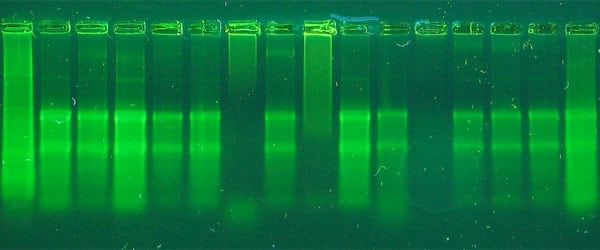
2. Problem: Degraded RNA/ low integrity
The rRNA bands appear smeared on a gel or the 18s is more intense than the 28s band. On the Agilent Bioanalyzer, you see a bigger 18s peak.
Cause: Degradation occurred at some point during processing. This can be difficult to pinpoint. It could have happened during collection and storage, or possibly during extraction. It could also have occurred post-isolation.
Solution: If the problem occurred during storage, make sure that samples are frozen immediately after collection. Use liquid nitrogen or -80°C storage. For animal tissue, use RNALater and then store at -20°C.
If the problem occurred during the extraction procedure, try adding beta-mercaptoethanol (BME) to the lysis buffer. Use 10 µl of 14.3 M BME per 1 ml of lysis buffer. BME will kill the RNases and help stabilize the sample during extraction.
If the sample is coming out of a freezer to be extracted and is not in a preservative solution, do not allow it to thaw. Homogenize it quickly in the presence of BME. Make sure not to leave behind any chunks of tissue. It needs to be completely lysed.
RNase degradation can also occur after isolation. Make sure that the water used for elution or resuspension of pellets is RNase free. Many kits provide water for RNA work that is DEPC treated or purified in other ways. More on the myths behind DEPC treatment and RNases can be read here.
In addition, 10 ways to work RNase free might also be a helpful article for you.
3. Problem: Inhibitors in the RNA
The RNA has an abnormally low 260/230 reading (below 1.0) or 260/280 reading or does not work in reverse transcription.
Cause: A low 260/230 in an RNA prep is indicative of guanidine salt carry over into the sample or organic inhibitors (such as humic acids or polysaccharides if the sample is environmental). Guanidine salts are used in TRIzol and in silica preps. These salts inactivate RNases, but will also inhibit proteins such as RT enzymes if present in the final RNA. A low 260/280 measurement indicates protein contamination.
Solution: For low 260/230 readings, the best approach is to try more washes of the RNA sample. If this is a TRIzol precipitate, try washing it with ethanol to desalt it. For silica preps, a few extra washes with 70-80% ethanol should clear the column of salts. For samples already purified that are not clean, try ethanol precipitation of the RNA to desalt.
For other inhibiting compounds, such as humic acid and polysaccharides, you may need to re-purify the sample on another column and wash it well before elution. Some of these compounds will not be removed from RNA (or DNA) with conventional methods because they are too similar to the nucleic acids. In this case, you may want to consider using inhibitor removal technology for environmental samples.
A low 260/280 reading from protein carry over most likely occurred because of using too much sample for the kit or method. The sample overwhelmed the chemistry and the proteins were not completely removed. Try cleaning up the sample with another round of your method- either phenol:chloroform and precipitate or add the binding salts and ethanol and bind to another silica spin filter. The protein should be easily removed. Next time, try using less sample and make sure homogenization is complete.
4. Problem: Low yields of RNA
The yield of RNA is lower than expected- either based on your previous results, or, based on reported yields for a certain tissue or cell type. RNA yields can vary greatly between different cultured cell types and in different tissues. For blood RNA, it can vary from person to person.
Cause: If the yield of RNA is lower than you expected or know it should be, and the RNA is intact (read: not degraded) , then the homogenization may not have been complete. To isolate RNA, a strong lysis is key. Tissues stored in RNALater will tend to be a little more difficult to homogenize. Low yields could be caused by mistakes in weighing of tissue or in the cell counts for cultured cells. You may have less cells than you think. With blood RNA, the buffy coat can vary based on your skill in collecting the white cell layer and each individual patient.
Solution:For cases where the RNA yield is low but the RNA is intact, focus on the homogenization method and make sure you are getting good shearing of the genomic DNA and release of RNA from all cells. If you see any pieces of tissue or debris in your homogenate, that is lost RNA.
It can be difficult to weigh small pieces of tissue so make sure you are using a scale accurate to the weight you need or you may see variation from prep to prep as well as other problems with purity and clogging columns when too much is used. For cells, it is important to have an accurate count if you want to have consistency.
If the RNA looks degraded, in addition to the yield being low, the problem could be that the homogenization was too hard and the sample was heated for too long. Try homogenizing in bursts of 30-45 seconds with 30 seconds of rest. Make sure to cut your tissue section and get it into cold guanidine lysis buffer (or TRIzol) quickly to prevent RNase activity during set up.
With silica spin filters, make sure the elution from the column is in enough volume to release the RNA from the membrane. More water will elute more RNA so use the largest volume that allows the RNA to be as concentrated as you need. Don’t try to use less volume than recommended by the manufacturer or you probably are leaving your RNA on the membrane. It is better to elute with more and concentrate after using ethanol precipitation, if maximal recovery is key.
And finally…
Isolation of RNA follows a similar protocol regardless of the sample source. For all samples, homogenization is the first step and the most important step. A good homogenization needs to break cells quickly to inactivate RNases in the lysis buffer, and needs to break genomic DNA down in size to make removal more efficient.
Republished June 2019.
65 Comments
Leave a Comment
You must be logged in to post a comment.
About the effect and cause of a low 260/230 ratio, I found an application note from Qiagen mentioning that the presence of guanidine salts in RNA is most often the cause of a low 260/230 ratio in RNA preparations made with TRIzol but has no influence in downstream applications (up to 100 mM guanidine thiocyanate (GITC) does not affect Ct values in RT-PCR). For instance, 260/230 ratios of about 1-1.5 indicate GITC concentrations of about 0.1-1 mM, so one must really push to get GITC concentrations that become a problem.
Source: Effects of low A260/A230 ratios in RNA preparations on downstream applications (https://www.qiagen.com/de/resources/download.aspx?id=11226191-0a82-4a9b-ba4a-99800b6f8595&lang=en)
Hi, it is very helpful, what kind of guanidine salts is the best for this application.could you tell us ,thanks!
i performing RNA extraction from freeze dryed human biopsies and i obtain:g concentration between 600 and 900 ng/microlit and 260/280 raatio is 2..14 to 3.0 but on gel I am not able to see 2 bands .. plz suggest