We received the following question from Bitesize Bio reader, Beheroze Sattha. It relates to a problem with absolute quantification using plasmids for standard curves. Since many people use this technique it is an interesting one question for us to explore, and it also gives us a great opportunity to cover some important tips for performing qPCR with a new template for standard curves.
Question:
I would like your help to assign copy numbers to plasmid standard dilutions. I cloned a portion of the mitochondrial D-loop gene and after plasmid prep of a single clone made serial dilutions (1:10). In my lab previously they would do a lightcycler run on those serial dilutions using SYBR Green and then assign copy numbers based on the crossing points. The theory used was that a crossing point (CP) of 32.7 would be equal to 10 copies. So the dilution closest to CP of 32.7 would be 10 copies with increasing 10 fold copies for the earlier dilutions (because I had made 1:10 serial dilutions: 1:10, 1:100, 1:1000, etc.
Then I learned from a co-worker that it is better to use the following website:
https://www.uri.edu/research/gsc/resources/cndna.html
Choose a free resource to help you move forward
PROTOCOL
Chemically Competent Cells Protocol
DOWNLOAD
Nanodrop Results Interpretation Card
So I entered the nanodrop reading of the undiluted plasmid standard and the length of template (TOPO vector and length of my insert) and got the copy number.
The problem is that there is a 10 fold difference between these two methods. 1:1000 dilution of the plasmid standard gives me 8.44E7 copies using the website and 9.99E6 using lightcycler CP.
******
Thanks for your question!
There are a couple things to keep in mind here and I think you’ll be able to solve this problem.
1- The standard curve results and crossing point or Cq numbers are not going to be identical every time. You didn’t mention whether the original standard curve data was performed with the cloned gene or with gDNA or with the PCR product for the gene itself. If the original standard curves were determined with one type of template and now you have a plasmid template, there will be a difference. Even a change of 1 cycle can have a big effect in absolute quantification.
2- Have you checked the efficiency of the plasmid template? How does it compare to the efficiency of the template used to generate the previous standard curve? If they are different, the quantification will be different. Ideally you want the efficiency to be above 90%. If the efficiency is below 80%, you won’t want to use this data and you may need to redesign the primers or optimize the chemistry or running times.
2- Every time you order new primers or a new enzyme kit (of a different lot#), you will want to repeat the standard curve results because the numbers can shift. As long as PCR efficiency is high, the data will be accurate, but the Cq may very well be different with new reagents.
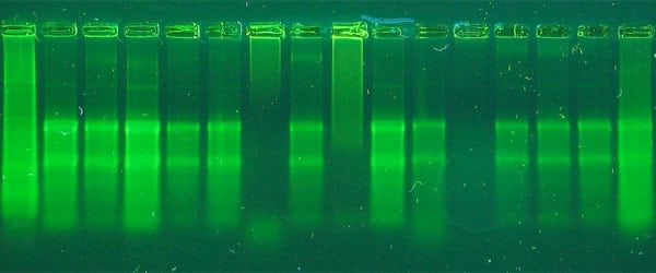
3- If you used a plasmid for a standard curve, did you linearize it for qPCR? Many people report that using supercoiled plasmid for standards can cause some variance in results. Try linearizing it first. Here is a recent publication on the subject.
4- When calculating the copy numbers, you may use the length of the PCR amplicon or the entire plasmid. When using the website above, if you use the size of just the amplicon as your input, make sure to adjust the amount of DNA going in to reflect the proportion of the plasmid (see the example in the comments). If you do not, your reading will be 10 fold off.
5- Make sure your negative controls are negative. Working with plasmids can be tricky because they can easily contaminate solutions. Make sure you have negative controls that are not amplifying because this will boost the real samples and result in inaccurate quantification.
6- Always do the melt curve analysis when using SYBR green and make sure you amplified a single product. Amplification from dimers will add fluorescence and result in an artificially low Cq. You can remedy this by performing an extra data acquisition step at a temperature above where the dimers melt and below where the real product melts. Alternatively, you may want to redesign the primers if dimer formation occurs even in samples with the highest amount of plasmid.
7- With plasmids, it is easy to overload the reaction and have Cq values so early that the detection won’t be accurate. Some instruments have a pre-set baseline setting where they subtract any fluorescent signals generated too early, assuming it is background noise. If you have too much signal in cycles 1-10, this can happen. You don’t want to have samples coming up early so dilute until the first sample has a Cq of 15-18. The subtraction of strong fluorescence in the early cycles will cause all of the data from the more dilute samples to shift right, causing later Cq values than what they are.
Summary:
When using a new plasmid as a standard in qPCR, do an efficiency check first and compare to the efficiency of the previous assay. For best results, an assay needs to have >90% efficiency, although there are formulas you can use that normalize for differences in efficiency. It is not uncommon that the Ct values are not exactly the same from user to user and from assay to assay for the same gene but with different primers. Just make sure that when calculating copy numbers, you are using the length of the template being amplified and not the entire plasmid which is probably 30 fold bigger than the template and could be the cause of the 10 fold difference in copy number results between the two methods you are comparing. Finally, it’s always good to re-check your standard curve with each new purchase of reagents to make sure no new variables are introduced that could throw off the quantification and make months of work unusable.
You made it to the end—nice work! If you’re the kind of scientist who likes figuring things out without wasting half a day on trial and error, you’ll love our newsletter. Get 3 quick reads a week, packed with hard-won lab wisdom. Join FREE here.